. 2013 May 1;41(10):5413-27.
doi: 10.1093/nar/gkt186. Epub 2013 Apr 4.

The monomeric GIY-YIG homing endonuclease I-BmoI uses a molecular anchor and a flexible tether to sequentially nick DNA

Affiliations

The monomeric GIY-YIG homing endonuclease I-BmoI uses a molecular anchor and a flexible tether to sequentially nick DNA

Benjamin P Kleinstiver et al. Nucleic Acids Res. .

Abstract

The GIY-YIG nuclease domain is found within protein scaffolds that participate in diverse cellular pathways and contains a single active site that hydrolyzes DNA by a one-metal ion mechanism. GIY-YIG homing endonucleases (GIY-HEs) are two-domain proteins with N-terminal GIY-YIG nuclease domains connected to C-terminal DNA-binding and they are thought to function as monomers. Using I-BmoI as a model GIY-HE, we test mechanisms by which the single active site is used to generate a double-strand break. We show that I-BmoI is partially disordered in the absence of substrate, and that the GIY-YIG domain alone has weak affinity for DNA. Significantly, we show that I-BmoI functions as a monomer at all steps of the reaction pathway and does not transiently dimerize or use sequential transesterification reactions to cleave substrate. Our results are consistent with the I-BmoI DNA-binding domain acting as a molecular anchor to tether the GIY-YIG domain to substrate, permitting rotation of the GIY-YIG domain to sequentially nick each DNA strand. These data highlight the mechanistic differences between monomeric GIY-HEs and dimeric or tetrameric GIY-YIG restriction enzymes, and they have implications for the use of the GIY-YIG domain in genome-editing applications.


Figures

Figure 1.
Figure 1.
I-BmoI interactions with substrate and monomeric cleavage models. (A) Schematic of the modular structure of I-BmoI interacting with intronless thyA substrate. Bottom- and top-strand nicking sites are indicated by filled and open triangles, respectively. IS, intron-insertion site. NUMOD, nuclease-associated modular DNA-binding domains. (B) Models for DSB formation by monomeric nucleases with single active sites. For transient dimerization, the secondary enzyme molecule can dimerize with the primary DNA-bound molecule from solution or via synapsis as a substrate-bound molecule at an additional target site.
Figure 2.
Figure 2.
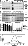
Identification of stable I-BmoI domains. (A) Images of Coomassie-stained SDS-gels of trypsin-limited proteolysis time-course experiments performed on I-BmoI and I-BmoI pre-incubated with intronless thyA substrate. Aliquots were removed at the indicated time points. In-gel trypsin digest followed by mass spectrometry was performed to identify the peptide products indicated with an asterisk. M, protein marker (sizes in kDa indicated to the left). (B) (top panel) Coomassie-stained SDS-gel of trypsin-limited proteolysis time-course experiments of I-BmoI in complex with intronless or intron-containing substrates. (bottom panel) Plot of the fraction of full-length I-BmoI remaining or increase of the catalytic domain peptide over time. Fraction of total protein is calculated after normalization to the untreated lane, with error bars representing the standard deviation of two replicates. (C) Enthalpy of transition curves for differential scanning calorimetry of I-BmoI or I-BmoI pre-incubated with substrate.
Figure 3.
Figure 3.
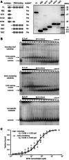
I-BmoI domains have distinct affinities for DNA. (A) Schematic of I-BmoI truncations and image of a Coomassie-stained SDS-page gel containing 1.5 µg of each soluble construct (left and right panels, respectively). FL, full-length I-BmoI; N92, residues 1–92 of I-BmoI; N111, 1–111; N130, 1–130; N154, 1–154; 92C, 92–266; 106C, 1–266; and 130C, 130–266. (B). Images of native gel-shift experiments for N154-binding reactions performed on labeled intronless, intron-containing and non-specific substrate. The left lane contains substrate only, and lanes 1 through 16 contain serial dilutions of N154 from 52.3 μM to 1.60 nM. (C) Graph of the binding curves of N154 I-BmoI on intronless thyA or intron-containing substrate, with data from three replicates plotted (see also Supplementary Table S3).
Figure 4.
Figure 4.
I-BmoI is a monomer in solution and in complex with thyA substrate. Graph of gel filtration elution profile of I-BmoI, The 46mer thyA substrate, or I-BmoI:substrate complex, with observed molecular weights indicated. Standards used to generate the elution volume standard curve are shown. Gel filtration analyses were performed in duplicate. Ve, elution volume; Vo, void volume.
Figure 5.
Figure 5.
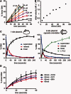
I-BmoI functions as a monomer. (A) Graph of initial reaction progresses for cleavage assays with eight I-BmoI concentrations expressed as per cent linear product. (B) Plot of initial reaction velocity versus I-BmoI concentration. (C) Graph of reaction progress for cleavage assays with I-BmoI and one- or two-site substrate plasmids (left and right panels, respectively). L1 + L2, linear products from two-site plasmid cleavage. (D) Domain addition experiments. Graph of linear product formation for cleavage assays with 22 nM I-BmoI and 10 nM plasmid. Reactions were supplemented with 500-fold molar excess of N111, N130 or BSA. All cleavage assays were performed in triplicate.
Figure 6.
Figure 6.
I-BmoI does not function via an intra-strand hairpin mechanism. Image of I-BmoI-binding reactions performed with dual-labeled thyA substrate (WT), bottom-strand pre-nicked substrate between −5/−4 (−4 nick) and bottom-strand pre-nicked substrate lacking the 3′-OH at the nick site (−4 ddGTP). Reactions were performed with 10 mM EDTA or 10 mM MgCl2. Schematic and naming of complexes are as previously described (34). Reactions were performed in duplicate showing similar results.
Figure 7.
Figure 7.
Insertions in the I-BmoI substrate spacer reduce or abolish cleavage activity. (A) Schematic of the wild-type thyA I-BmoI substrate and the substrates generated containing three- or five-nucleotide insertions. Filled and open triangles represent the bottom- and top-strand nicking sites, respectively. (B) Progress curves for cleavage reactions on thyA, Ins1 and Ins2 substrates in 10 mM MgCl2. Curves are generated from reactions performed in triplicate.
Figure 8.
Figure 8.
An in vivo screen identifies I-BmoI nucleotide preference at G + 7. (A) (top) Schematic representation of the randomized cleavage site from −6 to +8 with G − 2 fixed (in bold enlarged font). (bottom) The pKoxRCS library and I-BmoI expression plasmids. Filled and open triangles represent the bottom- and top-strand nicking sites, respectively. (B) Work flow to select for cleavable target sites. (C) Sequencing results from cleavable targets represented as the difference in nucleotide proportion from the clones in the survivor pool versus the input library.
Figure 9.
Figure 9.
Mutations at G + 7 affect I-BmoI activity. (A) Plot of reaction progress with I-BmoI and G + 7 (WT) or G + 7T plasmid substrates in 0.5 mM MgCl2. Curves are plotted as a fit to the mean of three replicates with standard deviation shown. (B) In vivo survival of I-BmoI on thyA, G + 7A, G + 7C and G + T toxic reporter plasmid substrates, with four replicates plotted. (C) Denaturing gel image of in-gel OP-Cu footprinting reactions with R27A I-BmoI on bottom-strand–labeled G + 7 (WT), G + 7A, G + 7C and G + 7T substrates (Unb, unbound substrate; UC, full-length I-BmoI bound to substrate). Sites that are hypersensitive to the footprinting reagent are highlighted with an asterisk, and filled triangles indicate the bottom-strand nick site. To the right of the gel image is a graph of the normalized pixel density ratio for bands in the UC lane versus the Unb lane. (D) Coomassie-stained SDS-gel of trypsin-limited proteolysis time-course experiments of I-BmoI in the presence of G + 7 (WT), G + 7A, G + 7C and G + 7T substrates. (E) Plot of the fraction of full-length I-BmoI remaining over time for reactions shown in panel (D), calculated as the fraction remaining after normalization to the untreated lane (error bars represent the standard deviation of two replicates).
Figure 10.
Figure 10.
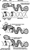
The domains of I-BmoI form distinct DNA-contacts to distort DNA before cleavage. (A) Schematic of I-BmoI domains and substrate modularity. The protease-resistant domains of I-BmoI and substrate bases required for efficient cleavage are highlighted. Bottom- and top-strand nicking sites are indicated by filled and open triangles, respectively. (B) Model of I-BmoI-bending DNA to induce substrate distortions near the bottom-strand nick site (indicated by white and grey stars that represent OP-Cu hypersensitivity for R27A and WT I-BmoI footprinting reactions, respectively). Rotation of the enzyme:substrate complex after bottom-strand nicking to reposition the nuclease domain for top-strand nicking is indicated by a dashed arrow around a vertical axis. Interactions at G − 2 and G + 7 may form a hinge necessary to distort DNA. (C) Schematic of nucleotide substitutions and insertions that affect I-BmoI catalytic activity.

Similar articles

Cited by

References

    1. Yang W. Nucleases: diversity of structure, function and mechanism. Q. Rev. Biophys. 2011;44:1–93. - PMC - PubMed
    1. Stoddard BL. Homing endonuclease structure and function. Q. Rev. Biophys. 2005;38:49–95. - PubMed
    1. Galburt EA, Stoddard BL. Catalytic mechanisms of restriction and homing endonucleases. Biochemistry. 2002;41:13851–13860. - PubMed
    1. Taylor GK, Stoddard BL. Structural, functional and evolutionary relationships between homing endonucleases and proteins from their host organisms. Nucleic Acids Res. 2012;40:5189–5200. - PMC - PubMed
    1. Kim YG, Cha J, Chandrasegaran S. Hybrid restriction enzymes: zinc finger fusions to fok I cleavage domain. Proc. Natl Acad. Sci. USA. 1996;93:1156–1160. - PMC - PubMed

Publication types

MeSH terms