. 2013 May;41(9):4976-87.
doi: 10.1093/nar/gkt182. Epub 2013 Apr 4.

LncRNA loc285194 is a p53-regulated tumor suppressor

Affiliations

LncRNA loc285194 is a p53-regulated tumor suppressor

Qian Liu et al. Nucleic Acids Res. 2013 May.

Abstract

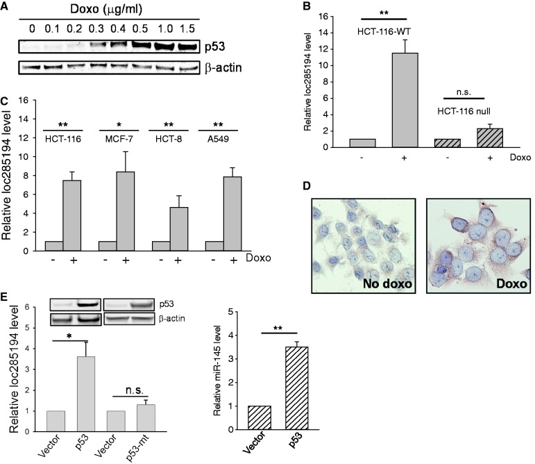
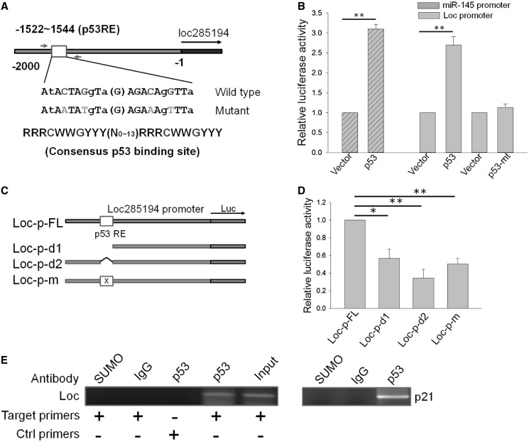
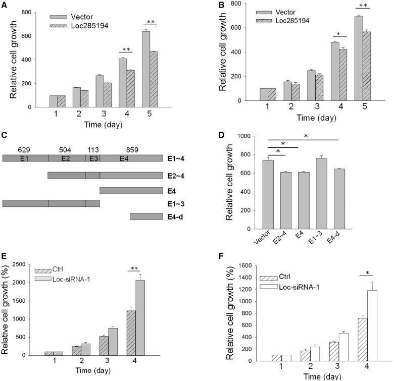
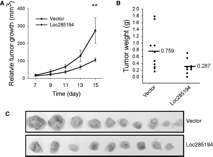
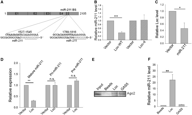
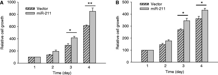
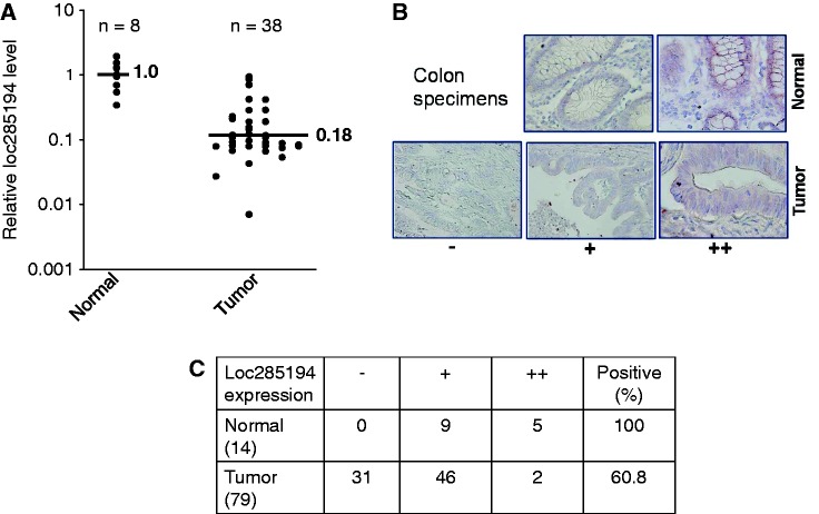
Protein-coding genes account for only a small part of the human genome, whereas the vast majority of transcripts make up the non-coding RNAs including long non-coding RNAs (lncRNAs). Accumulating evidence indicates that lncRNAs could play a critical role in regulation of cellular processes such as cell growth and apoptosis as well as cancer progression and metastasis. LncRNA loc285194 was previously shown to be within a tumor suppressor unit in osteosarcoma and to suppress tumor cell growth. However, it is unknown regarding the regulation of loc285194. Moreover, the underlying mechanism by which loc285194 functions as a potential tumor suppressor is elusive. In this study, we show that loc285194 is a p53 transcription target; ectopic expression of loc285194 inhibits tumor cell growth both in vitro and in vivo. Through deletion analysis, we identify an active region responsible for tumor cell growth inhibition within exon 4, which harbors two miR-211 binding sites. Importantly, this loc285194-mediated growth inhibition is in part due to specific suppression of miR-211. We further demonstrate a reciprocal repression between loc285194 and miR-211; in contrast to loc285194, miR-211 promotes cell growth. Finally, we detect downregulation of loc285194 in colon cancer specimens by quantitative PCR arrays and in situ hybridization of tissue microarrays. Together, these results suggest that loc285194 is a p53-regulated tumor suppressor, which acts in part through repression of miR-211.


Figures

Figure 1.
Figure 1.
Identification of loc285194 as a p53-induced lncRNA. (A) Detection of p53 induction in response to doxo by western blot. (B) Effect of doxo on loc285154 expression in HCT-116-WT and HCT-116 null cells. The cells were treated with doxo at 1 µg/ml for 24 h before extraction of total RNA for qRT-PCR. Error bars represent SEM, n = 3. **P < 0.01; n.s., not significant. (C) Induction of loc285194 in HCT-116, MCF-7, HCT-8 and A549 cells in response to doxo. The cells were treated the same way as in (B). Error bars represent SEM, n = 3. **P < 0.01; *P < 0.05. (D) Induction of loc285194 in doxo-treated cells as detected by ISH. (E) Induction of loc285194 by ectopically expressed p53. HCT-116 WT cells were first transfected with vector or wild-type p53 or mutant p53 (R175H) overnight; their expression was confirmed by western blot (top panel). After changing medium, the cells were allowed to further grow for 24 h before extraction of total RNA (bottom panel). Error bars represent SEM, n = 3. *P < 0.05.
Figure 2.
Figure 2.
p53 induces loc285194 by direct interacting with loc285194 promoter. (A) Schematic description of loc285194 promoter with a potential p53 response element (p53RE). Also shown are sequences for mutant p53RE and p53RE consensus. Relative position of primers used for ChIP assay (E) was indicated by arrows. (B) Induction of loc285194 promoter activity by wild-type p53, but not mutant p53. HCT-116 WT cells were transfected with loc285194 luciferase reporter (loc-p-FL) along with vector, wild-type p53 or mutant p53 (R175H). The cells were harvested for luciferase assays 24 h after transfection. The miR-145 promoter reporter served as a positive control. Error bars represent SEM, n = 3. **P < 0.01. (C) Deletion analysis of the promoter activity to determine the role of the potential p53RE in p53 regulation of loc285194. Loc-p-FL is a full-length promoter; Loc-p-d1 and Loc-p-d2 carry a deletion involving p53RE; Loc-p-m is a mutant at p53RE, as indicated in Figure 2A. (D) Relative luciferase activity for corresponding constructs in Figure 2C. Luciferase assay was conducted as in Figure 2B. Error bars represent SEM, n = 3. **P < 0.01; *P < 0.05. (E) Confirmation of p53 binding to p53RE in loc285194 promoter as detected by ChIP assay. Control primers Loc285194-ChIP-Ctrl-5.1 and Loc285194-ChIP-Ctrl-5.1 were derived from outside of p53RE. Immunoglobulin G and SUMO antibody were used as negative controls; p21 served as a positive control.
Figure 3.
Figure 3.
Loc285194 inhibits tumor cell growth in vitro. (A) Inhibition of cell growth by loc285194 in HCT-116 WT cells. The cells were first transfected with vector or loc285194 overnight and then split into 12-well plates. Cell number was counted from day 1 to day 5 by trypan blue method. Error bars represent SEM, n = 3. **P < 0.01. (B) Inhibition of cell growth by loc285194 in MCF-7 cells. The experiment was conducted the same way as in Figure 3A. Error bars represent SEM, n = 3. **P < 0.01; *P < 0.05. (C) Identification of the minimal region of loc285194 capable of inhibiting cell growth. Deletion constructs were made by PCR using primers (Supplementary Table S1) and then cloned into pCDH-MSCV-EF1-GFP-T2A-Puro. The length of each of 4 exons for loc285194 is indicated by number of nucleotides above E1∼4. E1∼4 is consisted of all 4 exons; E2∼4 is consisted of exon 2, 3 and 4; E1∼3 is consisted of exon 1, 2 and 3; E4 carries exon 4 only; and E4-d carries a deletion of the first 247 nucleotides of exon 4. (D) Effect of loc285194 and its deletion constructs corresponding to Figure 3C on cell growth. The experiment was conducted in HCT-116 WT cells with the same way as in Figure3 A. Error bars represent SEM, n = 3. *P < 0.05. (E) and (F) Suppression of loc285197 by RNAi increases cell growth in HCT-116 WT (E) and HCT-116 null cells (F). Cells were first transfected with control siRNA or loc285192 siRNAs overnight and then split into 12-well plates. The cell number was counted from day 1 as 100% to day 4. Error bars represent SEM, n = 3. **P < 0.01; *P < 0.05.
Figure 4.
Figure 4.
Loc285194 inhibits tumor growth in a xenograft mouse model. (A) Tumor growth curve. HCT-116 WT cells were transfected with vector or loc285194 and then injected into nude mice as described in ‘Materials and Methods’ section. Tumor growth was measured from day 7 after injection of tumor cells. Error bars represent SEM, n = 10. **P < 0.01. (B) Tumor weight when tumors were harvested. Error bars represent SEM, n = 10. *P < 0.05. (C) Total number of tumors after removal from mice.
Figure 5.
Figure 5.
Reciprocal negative regulation of miR-211 and loc285194. (A) Loc285194 carries two miR-211 binding sites at exon 4. Alignment between them is shown underneath with miR-211 seed sequences underlined. (B) Suppression of miR-211 expression by loc285194. HCT-116 cells were first transfected with vector, wild-type loc285194 (Loc-WT) or mutant loc285194 (Loc-d) with a deletion of two miR-211 binding sites, and 24 h after transfection, total RNA was isolated for qRT-PCR. Error bars represent SEM, n = 3. **P < 0.01. (C) Effect of miR-211 on loc285194 expression. HCT-116 WT cells were transfected with vector or miR-211, and total RNA was isolated for qRT-PCR 24 h after transfection. Error bars represent SEM, n = 3. *P < 0.05. (D) Effect of loc285194 on mature miR-211, pri-miR-211 and pre-miR-211. Error bars represent SEM, n = 3. **P < 0.01; n.s., not significant. (E) Pulldown of Ago2 by biotin-labeled loc285194 or GAS5 RNA probe, as detected by western blot. The GAS5 lane was composed from the same gel with the same contrast. (F) Detection of miR-211 in the pellet precipitated by the loc285194 probe, but not in the pellet precipitated by the GAS5 probe. Error bars represent SEM, n = 3. **P < 0.01.
Figure 6.
Figure 6.
miR-211 promotes cell growth in HCT-116 WT (A) and MCF-7 cells (B). Cells were transfected with vector or miR-211 overnight, and then split into 12-well plates. Cell number was countered from day 1 (as 100%) to day 4. Error bars represent SEM, n = 3. **P < 0.01; *P < 0.05.
Figure 7.
Figure 7.
Loc285194 is downregulated in colon cancer specimens. (A) Detection of loc285194 in TissueScan arrays by quantitative PCR. To calculate relative expression of loc285194, we took the average value of eight normal tissue samples as one and then compared each value of tumor specimens to the average value of normal tissue. Among 40 tumor samples, we only detected loc285194 expression in 38 samples; the other two were undetectable, which could be also counted as downregulation, but were not included here. (B) and (C) Detection of loc285194 in colon cancer tissue microarrays by ISH as described in ‘Materials and Methods’ section. (B) Representative pictures showing signal intensity scale: ‘0’ = negative; ‘ +’ = weak positive; ‘++’ = strong positive. (C) Loc285194 is downregulated in tumors compared with normal tissue.

Similar articles

Cited by

References

    1. Hwang HW, Mendell JT. MicroRNAs in cell proliferation, cell death, and tumorigenesis. Br. J. Cancer. 2006;94:776–780. - PMC - PubMed
    1. Croce CM, Calin GA. miRNAs, cancer, and stem cell division. Cell. 2005;122:6–7. - PubMed
    1. Hammond SM. MicroRNAs as oncogenes. Curr. Opin. Genet. Dev. 2006;16:4–9. - PubMed
    1. Esquela-Kerscher A, Slack FJ. Oncomirs - microRNAs with a role in cancer. Nat. Rev. Cancer. 2006;6:259–269. - PubMed
    1. Gregory RI, Shiekhattar R. MicroRNA biogenesis and cancer. Cancer Res. 2005;65:3509–3512. - PubMed

Publication types