Review
. 2013 Apr;9(4):192-200.
doi: 10.1038/nrneurol.2013.36.

Acute and chronic traumatic encephalopathies: pathogenesis and biomarkers

Affiliations
Review

Acute and chronic traumatic encephalopathies: pathogenesis and biomarkers

Steven T DeKosky et al. Nat Rev Neurol. 2013 Apr.

Abstract

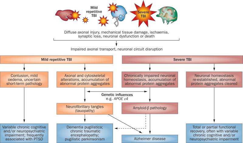
Over the past decade, public awareness of the long-term pathological consequences of traumatic brain injury (TBI) has increased. Such awareness has been stimulated mainly by reports of progressive neurological dysfunction in athletes exposed to repetitive concussions in high-impact sports such as boxing and American football, and by the rising number of TBIs in war veterans who are now more likely to survive explosive blasts owing to improved treatment. Moreover, the entity of chronic traumatic encephalopathy (CTE)--which is marked by prominent neuropsychiatric features including dementia, parkinsonism, depression, agitation, psychosis, and aggression--has become increasingly recognized as a potential late outcome of repetitive TBI. Annually, about 1% of the population in developed countries experiences a clinically relevant TBI. The goal of this Review is to provide an overview of the latest understanding of CTE pathophysiology, and to delineate the key issues that are challenging clinical and research communities, such as accurate quantification of the risk of CTE, and development of reliable biomarkers for single-incident TBI and CTE.


Figures

Figure 1
Figure 1
Spectrum of pathological features and outcomes of mild and severe TBI. Abbreviations: APOE, apolipoprotein E; PTSD, post-traumatic stress disorder; TBI, traumatic brain injury.
Figure 2
Figure 2
Histopathology of the temporal cortex in TBI and AD. Brain sections from ad | an individual with severe acute TBI and eh | a patient with AD are labelled for Aβ42 (a,e), Aβ40 (b,f), AβX–34 (c,g), or Apo-E (d,h). In the TBI case, Aβ42 plaques (a) are more abundant than Aβ40 plaques (b). Frequency of plaques with AβX–34 (c) is closer to that of Aβ40, whereas distribution of Apo-E-positive plaques (d) is similar to Aβ42 plaques. The AD temporal cortex shows profusion of all plaque types (e–h), as well as neurofibrillary tangles, neuropil threads, and vascular amyloidosis (g). Scale bar: 200 μm. Abbreviations: Aβ, amyloid-β; AD, Alzheimer disease; Apo-E, apolipoprotein E; TBI, traumatic brain injury. Permission obtained from Elsevier Ltd © Ikonomovic, M. D. et al. Exp. Neurol. 190, 192–203 (2004).
Figure 3
Figure 3
Histopathological features of chronic traumatic encephalopathy in a former professional football player. All sections are immunostained for abnormally phosphorylated tau using an AT-8 monoclonal antibody that detects hyperphosphorylated tau (Ser202 and Thr205). a | Scanning view of the hippocampus and parahippocampal cortex. Note intense immunostaining of the entire Ammon horn and subiculum, with focal involvement at the depths of sulci of the inferior temporal lobe. Original magnification ×1. b | Appearance of individual neurofibrillary tangles in the neocortex. Original magnification ×160. c | Neurofibrillary tangles in the anterior insular cortex form preferentially in the superficial layers (layers II–III), rather than in deeper layers (layers V–VI) as is more common in Alzheimer disease. Original magnification ×30. d | Tendency for perivascular tau deposition and neurofibrillary tangle formation in the frontal cortex. Original magnification ×60. Permission obtained from the American Medical Association © Shively, S. et al. Arch. Neurol. 69, 1245–1251 (2012).
Figure 4
Figure 4
18F-THK523 as a tauopathy marker. Autoradiography and microscopy analysis of hippocampal serial sections taken at autopsy from a 90-year-old patient with Alzheimer disease indicates that 18F-THK523 binds specifically to tau tangles, with no detectable binding to Aβ plaques. a | Low-magnification 18F-THK523 autoradiogram. bd | Higher-magnification microscopy and autoradiogram images of three serial sections immunostained with antibodies to tau (b), which label neurofibrillary tangles; with 18F-THK523 (c); or with antibodies to Aβ (d), which label Aβ plaques. 18F-THK523 labelling seems to colocalize with tau immunostaining of neurofibrillary tangles, but not with plaques. Abbreviation: Aβ, amyloid-β. Permission obtained from Oxford University Press © Fodero-Tavoletti, M. T. et al. Brain 134, 1089–1100 (2011).
Figure 5
Figure 5
CSF neurofilament light polypeptide in Olympic boxers after a bout. a | Individual CSF levels of neurofilament light polypeptide—a biomarker of axonal damage—in boxers after a bout and in age-matched controls. 25 of 30 (83%) of boxers had high CSF neurofilament light polypeptide levels, whereas 24 of 25 (96%) of controls had normal levels (below 125 ng/l), Wilcoxon signed rank test: P <0.001. b | Correlation between boxing exposure and CSF neurofilament light polypeptide levels. A lower score indicates fewer and easier fights, whereas higher scores indicate more, tougher fights with more head blows. Spearman's r = 0.40; P = 0.03. Abbreviation: CSF, cerebrospinal fluid. Figure is reproduced from Neselius, S. et al. PLoS ONE 7, e33606 (2012), which is published under an open-access license by the Public Library of Science.

References

    1. Centers for Disease Control and Prevention . Injury prevention and control: traumatic brain injury. Centers for Disease Control and Prevention; 2012. [online], http://www.cdc.gov/traumaticbraininjury/statistics.html.
    1. DeKosky ST, Ikonomovic MD, Gandy S. Traumatic brain injury—football, warfare, and long-term effects. N. Engl. J. Med. 2010;363:1293–1296. - PubMed
    1. US Department of Defense . Traumatic brain injury—Department of Defense special report. US Department of Defense; 2012. [online], http://www.defense.gov/home/features/2012/0312_tbi/
    1. Lange RT, Brickell TA, Ivins B, Vanderploeg R, French LM. Variable, not always persistent, postconcussion symptoms following mild TBI in U.S. military service members: a 5-year cross-sectional outcome study. J. Neurotrauma. http://dx.doi.org/10.1089/neu.2012.2743. - DOI - PubMed
    1. Wall PL. Posttraumatic stress disorder and traumatic brain injury in current military populations: a critical analysis. J. Am. Psychiatr. Nurses Assoc. 2012;18:278–298. - PubMed

Publication types