. 2013 Apr 5;340(6128):48-52.
doi: 10.1126/science.1229495.

A tissue-like printed material

Affiliations

A tissue-like printed material

Gabriel Villar et al. Science. .

Abstract

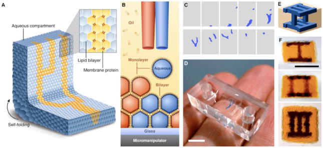
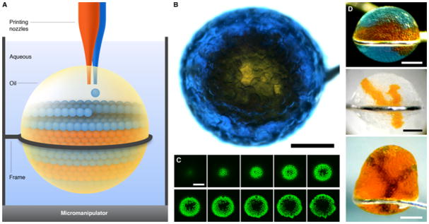
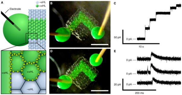
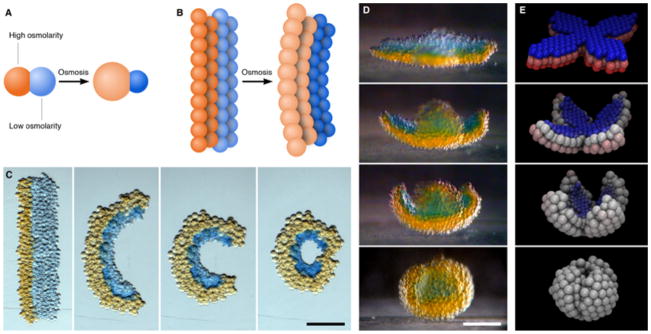
Living cells communicate and cooperate to produce the emergent properties of tissues. Synthetic mimics of cells, such as liposomes, are typically incapable of cooperation and therefore cannot readily display sophisticated collective behavior. We printed tens of thousands of picoliter aqueous droplets that become joined by single lipid bilayers to form a cohesive material with cooperating compartments. Three-dimensional structures can be built with heterologous droplets in software-defined arrangements. The droplet networks can be functionalized with membrane proteins; for example, to allow rapid electrical communication along a specific path. The networks can also be programmed by osmolarity gradients to fold into otherwise unattainable designed structures. Printed droplet networks might be interfaced with tissues, used as tissue engineering substrates, or developed as mimics of living tissue.


Conflict of interest statement

The authors declare no competing financial interests.

Figures

Fig. 1
Fig. 1
Printed droplet networks. (A) Illustration of a printed droplet network. (B) Schematic of the printing process. Two droplet generators eject droplets of different aqueous solutions into a solution of lipids in oil. The oil bath is mounted on a motorized micromanipulator. The droplets acquire a lipid monolayer, and form bilayers with droplets in the growing network. (C) Horizontal cross-sections of a design for a three-dimensional droplet network with a branching structure (blue) embedded in a cuboid (grey). The design comprises 20 layers of 50×35 droplets each; only alternate layers are shown. (D) Network printed according to the design in (C). Scale bar, 5 mm. (E) Schematic of a three-dimensional design that consists of 28 layers of 24×24 droplets each. (F) Three orthogonal views of a single network printed according to the design in (E). Scale bar, 1 mm.
Fig. 2
Fig. 2
Droplet networks printed in bulk aqueous solution. (A) Schematic of printing in aqueous solution. Aqueous droplets are ejected into a drop of oil suspended in bulk aqueous solution. Excess oil can be removed after printing by suction through a printing nozzle. (B) Micrograph of a network printed in aqueous solution, viewed from above. A core of orange droplets is surrounded by a shell of blue droplets, which contain the fluorescent dye pyranine. Scale bar, 400 μm. (C) Horizontal sections of the network in (B) obtained by confocal microscopy, showing the fluorescent shell of droplets around the non-fluorescent core. The sections span approximately the bottom 150 μm of the network. Scale bar, 400 μm. (D) Micrographs of three other networks printed in bulk aqueous solution. Scale bars, 400 μm.
Fig. 3
Fig. 3
Electrically conductive pathway. (A) Schematic of part of a network printed with an ionically conductive pathway. Only the green droplets and the large drop contain αHL pores. The large drop is impaled with an Ag/AgCl electrode. The magnified section illustrates the αHL pores in the bilayers around the αHL-containing droplets. (B) Photograph of a printed network with electrode-impaled drops placed on either end of the conductive pathway. The green droplets contain αHL, while the other droplets contain no protein. Scale bar, 500 μm. (C) Stepwise increase in the ionic current as measured in the configuration in (B), at 50 mV in 1 M KCl at pH 8.0. (D) Photograph of the network in (B), after separating one of the large drops and rejoining it onto the network away from the conductive pathway. Scale bar, 500 μm. (E) Selected portions of a single recording as measured in the configuration in (D) at 50 mV, showing transient increases in ionic current.
Fig. 4
Fig. 4
Self-folding droplet networks. (A) Schematic of two droplets of different osmolarities joined by a lipid bilayer. The flow of water through the bilayer causes the droplets to swell or shrink. (B) Schematic of a droplet network that comprises two strips of droplets of different osmolarities. The transfer of water between the droplets induces an overall deformation of the network. (C) Photographs of a rectangular network folding into a circle over ~3 h. The orange and blue droplets initially contain 250 mM KCl and 16 mM KCl, respectively. Scale bar, 250 μm. (D) Photographs of a flower-shaped network folding spontaneously into a hollow sphere. The orange and blue droplets initially contain 80 mM KCl and 8 mM KCl, respectively. The photographs cover a period of 8 h. Scale bar, 200 μm. (E) Frames from a folding simulation of a network with a similar initial geometry to the network in (D). Blue and red represent the lowest and highest initial osmolarities, respectively, and white indicates the average of the two.

Comment in

References

    1. Nakagawa S, Maeda S, Tsukihara T. Curr Opin Struct Biol. 2010;20:423. - PubMed
    1. Poulin P, Bibette J. Langmuir. 1998;14:6341.
    1. Funakoshi K, Suzuki H, Takeuchi S. Anal Chem. 2006;78:8169. - PubMed
    1. Malmstadt N, Nash MA, Purnell RF, Schmidt JJ. Nano Lett. 2006;6:1961. - PubMed
    1. Holden MA, Needham D, Bayley H. J Am Chem Soc. 2007;129:8650. - PubMed

Publication types