. 2013 Apr 5;340(6128):78-81.
doi: 10.1126/science.1234654.

Drosophila H1 regulates the genetic activity of heterochromatin by recruitment of Su(var)3-9

Affiliations

Drosophila H1 regulates the genetic activity of heterochromatin by recruitment of Su(var)3-9

Xingwu Lu et al. Science. .

Abstract

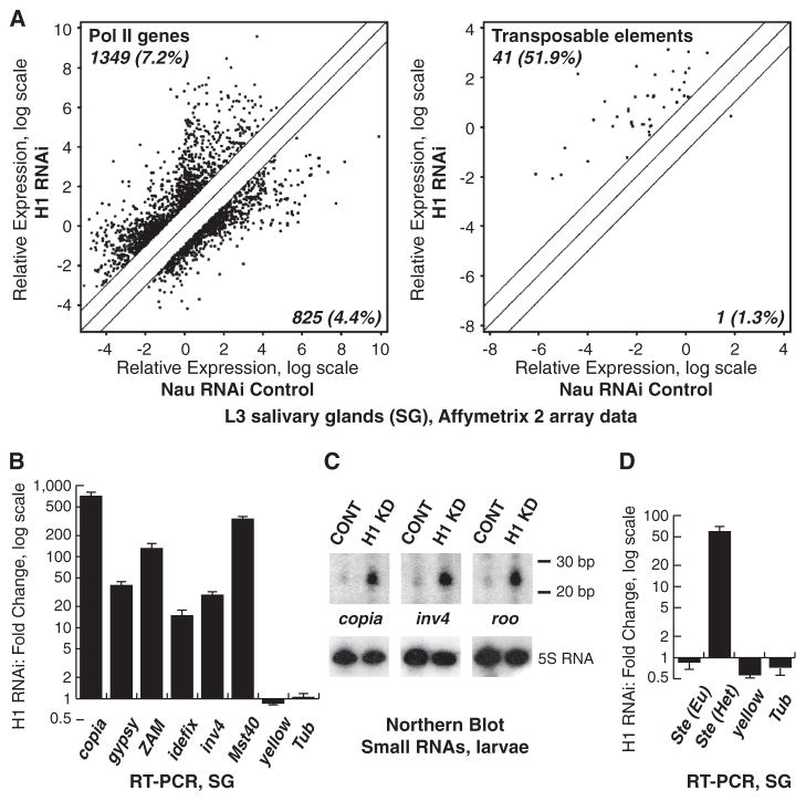
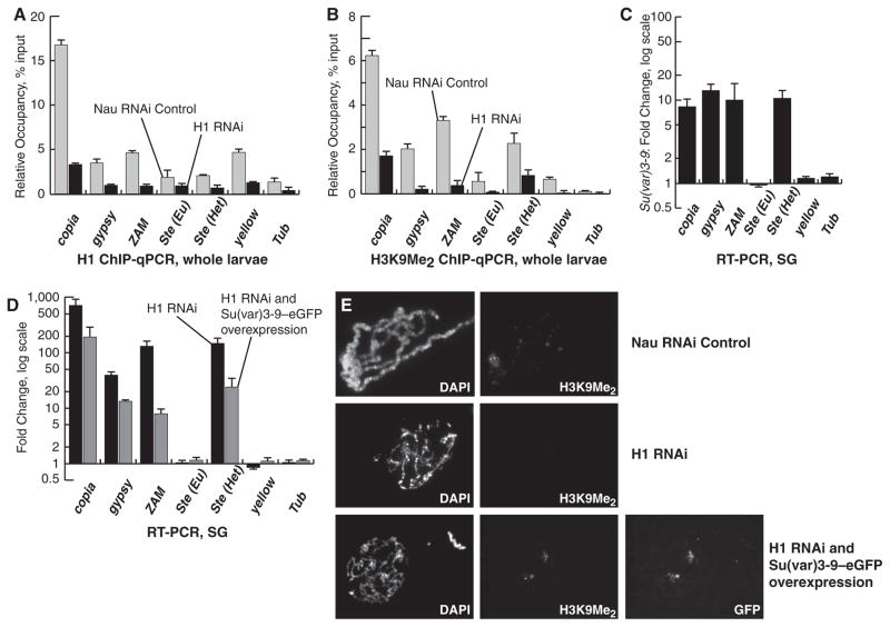
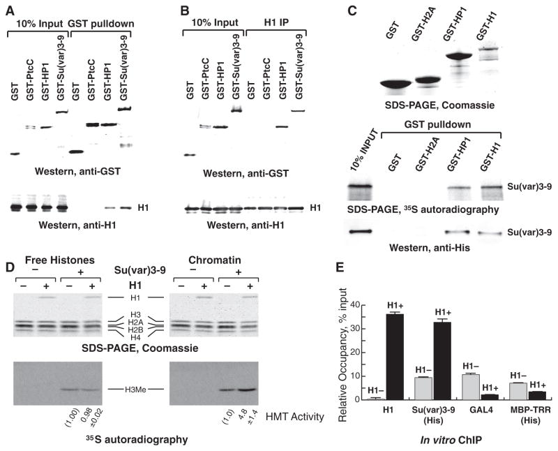
Eukaryotic genomes harbor transposable elements and other repetitive sequences that must be silenced. Small RNA interference pathways play a major role in their repression. Here, we reveal another mechanism for silencing these sequences in Drosophila. Depleting the linker histone H1 in vivo leads to strong activation of these elements. H1-mediated silencing occurs in combination with the heterochromatin-specific histone H3 lysine 9 methyltransferase Su(var)3-9. H1 physically interacts with Su(var)3-9 and recruits it to chromatin in vitro, which promotes H3 methylation. We propose that H1 plays a key role in silencing by tethering Su(var)3-9 to heterochromatin. The tethering function of H1 adds to its established role as a regulator of chromatin compaction and accessibility.


Figures

Fig. 1
Fig. 1. Drosophila H1 represses repetitive elements
(A) Transcript expression was examined by micro-array analyses in H1-depleted and control (Nau RNAi) SGs (4). Signal intensities are shown for transcripts in control (x axis) versus H1-depleted (y axis) samples. The diagonal lines indicate equal expression level or a twofold change. Significantly affected transcripts above or below twofold threshold are indicated by dots. (Left) Signals for protein-coding gene probes; (right) signals for probes annotated as TEs. Numbers in the top left and bottom right corners represent percentages of transcripts that are up- or down-regulated above threshold, relative to the total number of probes (18,833 protein coding genes, 79 TEs). (B) TE transcripts in SGs were analyzed by QRT-PCR. Fold changes were calculated as a ratio of signals for H1-depleted samples to those for control samples and normalized to RP49. Standard deviations are from triplicate PCR reactions for three independent experiments. (C) RNA was extracted from H1-depleted (H1 KD) and control (CONT) larvae and (top) analyzed by Northern blot with TE-specific probes. (Bottom) Hybridization with the 5S RNA probe (loading control). (D) QRT-PCR of transcripts for euchromatic (Eu) and heterochromatic (Het) copies of Ste was analyzed as in (B).
Fig. 2
Fig. 2. H1 represses repetitive elements in conjunction with Su(var)3-9
(A) The occupancy of H1 in larval chromatin was measured by qChIP. The ordinate indicates the amounts of qChIP DNA samples relative to input DNA. All experiments were performed in triplicate. Error bars, standard deviation. (B) The occupancy of the H3K9Me2 was measured by qChIP and presented as in (A). (C) QRT-PCR assays were performed in homozygous Su(var)3-9[6] and wild-type SGs. The data were analyzed as in Fig. 1B. (D) RNA was prepared from SGs from control, H1-depleted, and H1-depleted UAS:Su(var)3-9–eGFP larvae. QRT-PCR assays were performed as in (C). Black bars, H1-depleted SGs; gray bars, H1-depleted UAS:Su(var)3-9-eGFP SGs. (E) SGs from control (top), H1-depleted (middle), and H1-depleted UAS:Su(var)3-9–eGFP (bottom) larvae were dissected, and polytene spreads were stained with 4′,6′-diamidino-2-phenylindole (DAPI) and the indicated antibodies.
Fig. 3
Fig. 3. H1 physically interacts with and recruits Su(var)3-9 to chromatin in vitro
(A) GST fusion proteins were ectopically expressed in S2 cells and immunoprecipitated. The input and IP material was analyzed by immunoblotting with (top) GST- and (bottom) H1-specific antibodies. PtC, the C-terminal tail of Hedgehog receptor Patched (25) (negative control). (B) Reciprocal IP experiments with H1-specific antibody were performed as in (A). (C) Su(var)3-9 was expressed and 35S-labeled by in vitro translation in reticulocyte lysates or purified as a 6His-tagged protein from bacteria. GST fusion proteins were expressed in E. coli and incubated with Su(var)3-9. The pulled-down material was examined by SDS–polyacrylamide electrophoresis (SDS-PAGE) and Coomassie staining (top), autoradiography (middle), or 6His-specific antibody immunoblotting (bottom). (D) Free histones (left) or reconstituted chromatin (right) with and without H1 were incubated with radioactive S-adenosylmethionine (SAM) in the presence or absence of recombinant Su(var)3-9–His6. H3 methylation was examined by autoradiography (bottom) and corrected for H3 loading (top, Coomassie). H3 methylation was quantified in two independent experiments; the average and standard deviation are shown at the bottom. (E) In vitro reconstituted chromatin with (H1+, black bars) or without H1 (H1–, gray bars) was incubated with Su(var)3-9–His6, GAL4-VP16, or MBP-TRR–His6, crosslinked, and analyzed by in vitro QChIP. The ordinate indicates the amounts of qChIP DNA samples relative to input DNA. All experiments were performed in triplicate. Error bars, standard deviation.

References

    1. Wolffe AP. Chromatin: Structure and Function. Academic Press; San Diego: 1995.
    1. Elgin SC, Grewal SI. Curr Biol. 2003;13:R895. - PubMed
    1. Eissenberg JC, Reuter G. Int Rev Cell Mol Biol. 2009;273:1. - PubMed
    1. Lu X, et al. Genes Dev. 2009;23:452. - PMC - PubMed
    1. Vujatovic O, et al. Nucleic Acids Res. 2012;40:5402. - PMC - PubMed

Publication types

MeSH terms

Associated data