

Microengineered tumor models: insights & opportunities from a physical sciences-oncology perspective
- PMID: 23559404
- PMCID: PMC3714360
- DOI: 10.1007/s10544-013-9763-y
Microengineered tumor models: insights & opportunities from a physical sciences-oncology perspective
Abstract
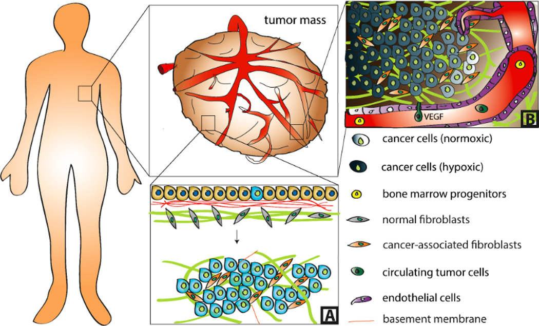
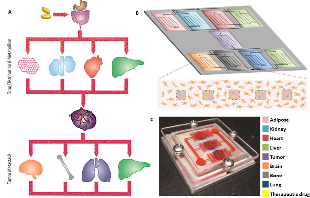
Prevailing evidence has established the fundamental role of microenvironmental conditions in tumorigenesis. However, the ability to identify, interrupt, and translate the underlying cellular and molecular mechanisms into meaningful therapies remains limited, due in part to a lack of organotypic culture systems that accurately recapitulate tumor physiology. Integration of tissue engineering with microfabrication technologies has the potential to address this challenge and mimic tumor heterogeneity with pathological fidelity. Specifically, this approach allows recapitulating global changes of tissue-level phenomena, while also controlling microscale variability of various conditions including spatiotemporal presentation of soluble signals, biochemical and physical characteristics of the extracellular matrix, and cellular composition. Such platforms have continued to elucidate the role of the microenvironment in cancer pathogenesis and significantly improve drug discovery and screening, particularly for therapies that target tumor-enabling stromal components. This review discusses some of the landmark efforts in the field of micro-tumor engineering with a particular emphasis on deregulated tissue organization and mass transport phenomena in the tumor microenvironment.
Conflict of interest statement
The authors declare that they have no conflict of interest.
Figures
Similar articles
-
Engineering strategies to capture the biological and biophysical tumor microenvironment in vitro.Adv Drug Deliv Rev. 2021 Sep;176:113852. doi: 10.1016/j.addr.2021.113852. Epub 2021 Jun 28. Adv Drug Deliv Rev. 2021. PMID: 34197895 Free PMC article. Review.
-
Microengineered 3D Tumor Models for Anti-Cancer Drug Discovery in Female-Related Cancers.Ann Biomed Eng. 2021 Aug;49(8):1943-1972. doi: 10.1007/s10439-020-02704-9. Epub 2021 Jan 5. Ann Biomed Eng. 2021. PMID: 33403451 Review.
-
In vitro models of tumor vessels and matrix: engineering approaches to investigate transport limitations and drug delivery in cancer.Adv Drug Deliv Rev. 2014 Apr;69-70:205-216. doi: 10.1016/j.addr.2013.11.011. Epub 2013 Dec 2. Adv Drug Deliv Rev. 2014. PMID: 24309015 Free PMC article. Review.
-
Engineering strategies to mimic the glioblastoma microenvironment.Adv Drug Deliv Rev. 2014 Dec 15;79-80:172-83. doi: 10.1016/j.addr.2014.08.012. Epub 2014 Aug 29. Adv Drug Deliv Rev. 2014. PMID: 25174308 Free PMC article. Review.
-
Microengineered cancer-on-a-chip platforms to study the metastatic microenvironment.Lab Chip. 2016 Oct 18;16(21):4063-4081. doi: 10.1039/c6lc00718j. Lab Chip. 2016. PMID: 27605305 Review.
Cited by
-
Gene Co-Expression Analysis Predicts Genetic Variants Associated with Drug Responsiveness in Lung Cancer.AMIA Jt Summits Transl Sci Proc. 2016 Jul 20;2016:32-41. eCollection 2016. AMIA Jt Summits Transl Sci Proc. 2016. PMID: 27570645 Free PMC article.
-
Three-dimensional in vitro tumor models for cancer research and drug evaluation.Biotechnol Adv. 2014 Nov 15;32(7):1256-1268. doi: 10.1016/j.biotechadv.2014.07.009. Epub 2014 Aug 10. Biotechnol Adv. 2014. PMID: 25116894 Free PMC article. Review.
-
The mechanical and pharmacological regulation of glioblastoma cell migration in 3D matrices.J Cell Physiol. 2019 Apr;234(4):3948-3960. doi: 10.1002/jcp.27209. Epub 2018 Aug 21. J Cell Physiol. 2019. PMID: 30132879 Free PMC article.
-
Functional biomaterials for biomimetic 3D in vitro tumor microenvironment modeling.In Vitro Model. 2023 Jan 27;2(1-2):1-23. doi: 10.1007/s44164-023-00043-2. eCollection 2023 Apr. In Vitro Model. 2023. PMID: 39872875 Free PMC article. Review.
-
A three-dimensional in vitro model of tumor cell intravasation.Integr Biol (Camb). 2014 Jun;6(6):603-10. doi: 10.1039/c3ib40170g. Integr Biol (Camb). 2014. PMID: 24763498 Free PMC article.
References
Publication types
MeSH terms
Grants and funding
LinkOut - more resources
Full Text Sources
Other Literature Sources
