

Role of (18)F-fluorodeoxyglucose Positron Emission Tomography scan in differentiating enhancing brain tumors
- PMID: 23559710
- PMCID: PMC3613621
- DOI: 10.4103/0972-3919.106698
Role of (18)F-fluorodeoxyglucose Positron Emission Tomography scan in differentiating enhancing brain tumors
Abstract
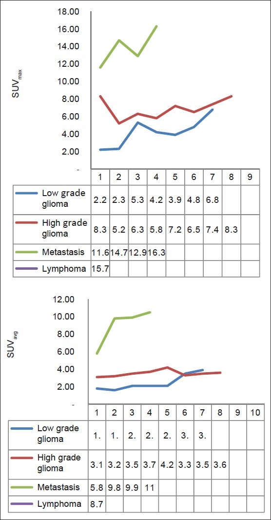
Aim: To determine whether F-18-fluorodeoxyglucose positron emission tomography (F-18-FDG PET) can be used to differentiate among common enhancing brain tumors such as gliomas, metastatic brain tumors, and lymphoma.
Materials and methods: We evaluated 20 patients with an enhancing brain tumor on magnetic resonance imaging (MRI). FDG PET scan was done in all patients pre operatively. For PET image analysis, regions of interest were placed over the tumor (T), contralateral cortex (C), and white matter (WM). Average and maximum pixel values were determined at each site. On the basis of these measurements, average and maximum standard uptake values (SUV avg and SUV max ) were calculated, and comparisons among lesions were then made.
Results: SUVavg and SUVmax are significantly higher for central nervous system (CNS) lymphoma than for other tumors (P < 0.01). High-grade gliomas showed significantly higher SUVavg and SUVmax than the low grade gliomas (P < 0.05) and metastatic tumor showed higher SUVavg and SUVmax than all gliomas, both low and high grade (P < 0.05). When the lowest values of CNS lymphoma parameter were used as cutoff levels to distinguish CNS lymphomas from other tumors (i.e. 100% sensitivity), SUVmax was the most accurate parameter. Using a SUVmax of 15.0 as a cutoff for diagnosing CNS lymphoma, only one case of metastasis (SUV max , 16.3) was found to be false positive in this study.
Conclusion: FDG PET appears to provide additional information for differentiating common enhancing malignant brain tumors, namely lymphoma versus high grade glioma and metastatic tumor, particularly when differential diagnoses are difficult to narrow using MRI alone.
Keywords: Brain tumor; F-18 fluorodeoxyglucose; PET; nuclear medicine.
Conflict of interest statement
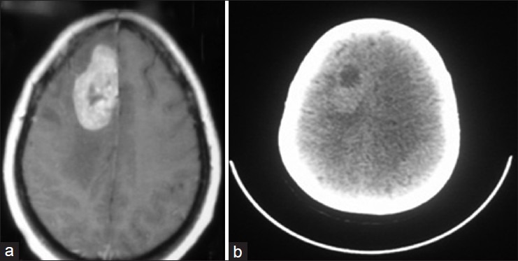
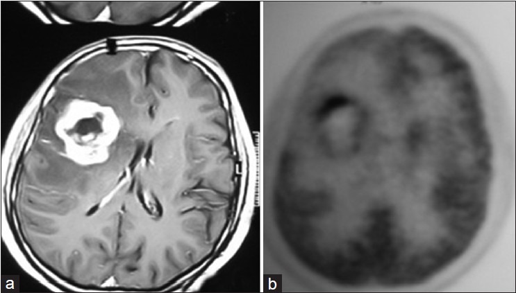
Figures
References
-
- Kosaka N, Tsuchida T, Uematsu H, Kimura H, Okazawa H, Itoh H. 18F-FDG PET common enhancing malignant brain tumors. AJR Am J Roentgenol. 2008;190:W365–9. - PubMed
-
- Calli C, Kitis O, Yunten N, Yurtseven T, Islekel S, Akalin T. Perfusion and diffusion MR imaging in enhancing malignant cerebral tumours. Eur J Radiol. 2006;58:394–403. - PubMed
-
- Hakyemez B, Erdogan C, Bolca N, Yildirim N, Gokalp G, Parlak M. Evaluation of different cerebral mass lesions by perfusion-weighted MR imaging. J Magn Reson Imaging. 2006;24:817–24. - PubMed
-
- Wahl RL. Nuclear medicine in clinical diagnosis and treatment. Vol. 2. New York: Churchill Livingstone; 1994. Positron emission tomography: Application in oncology; pp. 801–20.
