

Anterior and middle superior alveolar nerve block for anesthesia of maxillary teeth using conventional syringe
- PMID: 23559916
- PMCID: PMC3612188
- DOI: 10.4103/1735-3327.104870
Anterior and middle superior alveolar nerve block for anesthesia of maxillary teeth using conventional syringe
Abstract
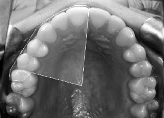
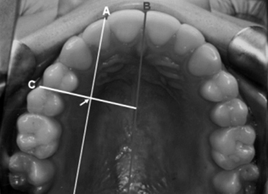
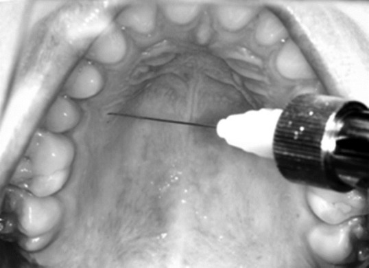
Background: Dental procedures in the maxilla typically require multiple injections and may inadvertently anesthetize facial structures and affect the smile line. To minimize these inconveniences and reduce the number of total injections, a relatively new injection technique has been proposed for maxillary procedures, the anterior and middle superior alveolar (AMSA) nerve block, which achieves pulpal anesthesia from the central incisor to second premolar through palatal approach with a single injection. The purpose of this article is to provide background information on the anterior and middle superior alveolar nerve block and demonstrate its success rates of pulpal anesthesia using the conventional syringe.
Materials and methods: Thirty Caucasian patients (16 men and 14 women) with an average age of 22 years-old, belonging to the School of Dentistry of Los Andes University, were selected. All the patients received an AMSA nerve block on one side of the maxilla using the conventional syringe, 1 ml of lidocaine 2% with epinephrine 1:100.000 was injected to all the patients.
Results: The AMSA nerve block obtained a 66% anesthetic success in the second premolar, 40% in the first premolar, 60% in the canine, 23.3% in the lateral incisor, and 16.7% in the central incisor.
Conclusions: Because of the unpredictable anesthetic success of the experimental teeth and variable anesthesia duration, the technique is disadvantageous for clinical application as the first choice, counting with other techniques that have greater efficacy in the maxilla. Although, anesthetizing the teeth without numbing the facial muscles may be useful in restorative dentistry.
Keywords: AMSA nerve block; dental anesthesia; local anesthesia; maxillary nerve.
Conflict of interest statement
Figures
Similar articles
-
Palatal Injection does not Block the Superior Alveolar Nerve Trunks: Correcting an Error Regarding the Innervation of the Maxillary Teeth.Cureus. 2018 Jan 28;10(1):e2120. doi: 10.7759/cureus.2120. Cureus. 2018. PMID: 29600124 Free PMC article. Review.
-
Effectiveness of Anterior and Middle Superior Alveolar Nerve Block for Anesthesia of Maxillary Teeth Using Conventional Syringe: A Randomized Prospective Study.J Maxillofac Oral Surg. 2022 Jun;21(2):616-619. doi: 10.1007/s12663-020-01432-w. Epub 2020 Aug 11. J Maxillofac Oral Surg. 2022. PMID: 35712390 Free PMC article.
-
A comparison of the anterior middle superior alveolar nerve block and infraorbital nerve block for anesthesia of maxillary anterior teeth.J Am Dent Assoc. 2010 Dec;141(12):1442-8. doi: 10.14219/jada.archive.2010.0106. J Am Dent Assoc. 2010. PMID: 21119128 Clinical Trial.
-
Alternative anesthetic technique for maxillary periodontal surgery.J Periodontol. 2008 Sep;79(9):1769-72. doi: 10.1902/jop.2008.070621. J Periodontol. 2008. PMID: 18771380
-
Current status of the anterior middle superior alveolar anesthetic injection for periodontal procedures in the maxilla.J Dent Anesth Pain Med. 2019 Feb;19(1):1-10. doi: 10.17245/jdapm.2019.19.1.1. Epub 2019 Feb 28. J Dent Anesth Pain Med. 2019. PMID: 30859128 Free PMC article. Review.
Cited by
-
Palatal Injection does not Block the Superior Alveolar Nerve Trunks: Correcting an Error Regarding the Innervation of the Maxillary Teeth.Cureus. 2018 Jan 28;10(1):e2120. doi: 10.7759/cureus.2120. Cureus. 2018. PMID: 29600124 Free PMC article. Review.
-
Pattern of buccal and palatal bone density in the maxillary premolar region: an anatomical basis of anterior-middle superior alveolar (AMSA) anesthetic technique.J Dent Anesth Pain Med. 2020 Dec;20(6):387-395. doi: 10.17245/jdapm.2020.20.6.387. Epub 2020 Dec 28. J Dent Anesth Pain Med. 2020. PMID: 33409367 Free PMC article.
-
Effectiveness of Anterior and Middle Superior Alveolar Nerve Block for Anesthesia of Maxillary Teeth Using Conventional Syringe: A Randomized Prospective Study.J Maxillofac Oral Surg. 2022 Jun;21(2):616-619. doi: 10.1007/s12663-020-01432-w. Epub 2020 Aug 11. J Maxillofac Oral Surg. 2022. PMID: 35712390 Free PMC article.
-
Effectiveness of anterior middle superior alveolar injection using a computer-controlled local anesthetic delivery system for maxillary periodontal flap surgery.J Dent Anesth Pain Med. 2019 Feb;19(1):45-54. doi: 10.17245/jdapm.2019.19.1.45. Epub 2019 Feb 28. J Dent Anesth Pain Med. 2019. PMID: 30859133 Free PMC article.
-
Effectiveness and Distribution of Anesthesia for a Modified Extra Oral Maxillo-Mandibular Nerve Block for Dento-Alveolar Procedures: A Prospective Cohort Study.J Maxillofac Oral Surg. 2024 Jun;23(3):561-567. doi: 10.1007/s12663-022-01755-w. Epub 2022 Jul 11. J Maxillofac Oral Surg. 2024. PMID: 38911425 Free PMC article.
References
-
- Blanton PC, Roda RS. The anatomy of local anesthesia. J Cal Dent Assoc. 1995;23:55–69. - PubMed
-
- Gomolka KA. The AMSA block: Local anesthesia without collateral numbness. CDS Rev. 2000;93:34. - PubMed
-
- Haas DA. An update on local anesthetics in dentistry. J Can Dent Assoc. 2002;68:546–51. - PubMed
-
- Friedman M, Hochman M. The AMSA injection: A new concept for local anesthesia of maxillary teeth using a computer-controlled injection system. Quintessence Int. 1998;29:297–303. - PubMed
LinkOut - more resources
Full Text Sources