

Management of polyostotic eosinophilic granuloma
- PMID: 23559966
- PMCID: PMC3612238
Management of polyostotic eosinophilic granuloma
Abstract
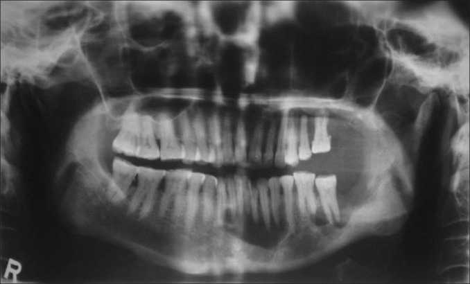
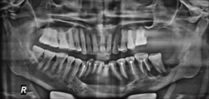
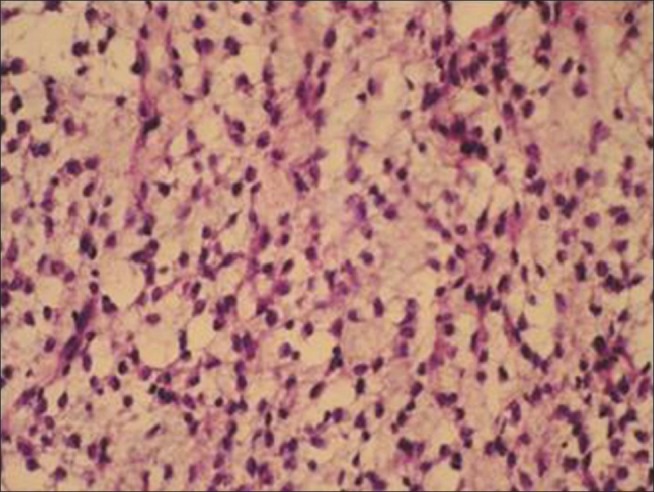
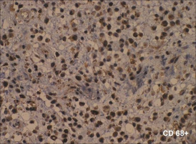
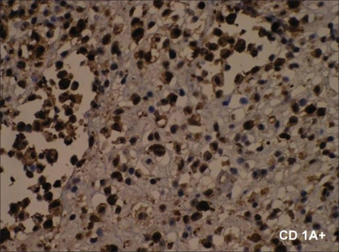
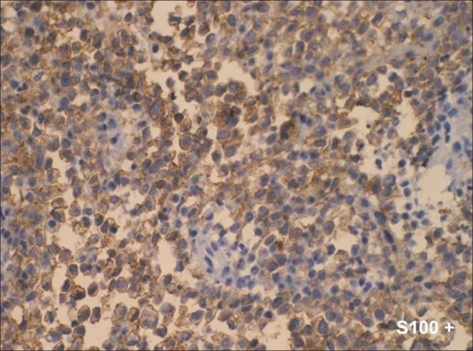
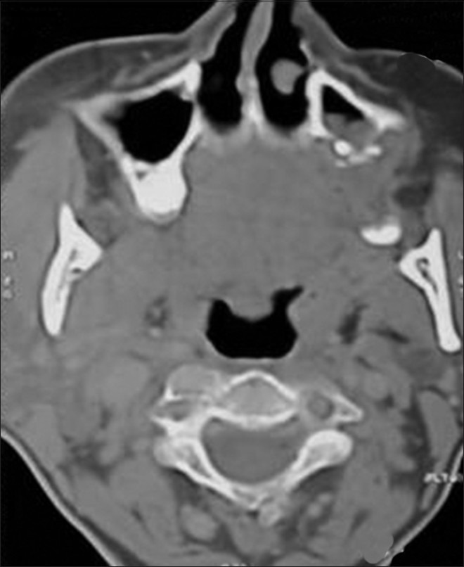
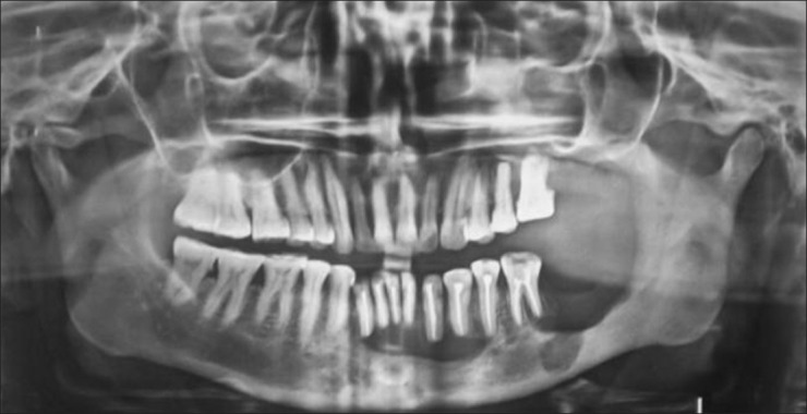
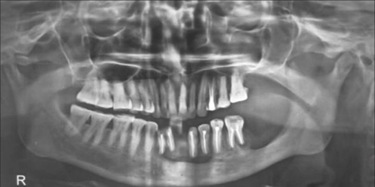
Eosinophilic granuloma is a rare disease which is difficult to diagnose clinically and radiographically. Localized Langerhans' cell histiocytosis, previously known as eosinophilic granuloma, mainly affects the skull, mandible, vertebrae, pelvis and ribs in children and the long bones of adults. We present a case report of a female who developed pain and swelling over the left mandibular region, and was later diagnosed as eosinophilic granuloma, which after administration of intralesional corticosteroid with surgical enucleation showed positive response. This disease is of importance to dental professionals because early clinical signs can occur in the jaw and can cause extensive destruction of the periodontal tissues and bone. The purpose of this case report is to describe a case of eosinophilic granuloma with emphasis on conservative approach for the treatment and the radiographic changes observed during and after the treatment.
Keywords: Adrenal cortex hormones; Langerhans’ cell histiocytosis; eosinophilic granuloma.
Conflict of interest statement
Figures
Similar articles
-
Eosinophilic Granuloma.2025 Jan 22. In: StatPearls [Internet]. Treasure Island (FL): StatPearls Publishing; 2025 Jan–. 2025 Jan 22. In: StatPearls [Internet]. Treasure Island (FL): StatPearls Publishing; 2025 Jan–. PMID: 32644464 Free Books & Documents.
-
Recurrent eosinophilic granuloma involving maxilla and mandible in an adult male: an unusual case report.Aust Dent J. 2021 Mar;66 Suppl 1:S88-S92. doi: 10.1111/adj.12861. Epub 2021 Jul 11. Aust Dent J. 2021. PMID: 34043826
-
Contribution to the radiological study of the eosinophilic granuloma of the mandible (unifocal granuloma due to Langerhans' cell histiocytosis).Radiol Med. 2005 Apr;109(4):414-20. Radiol Med. 2005. PMID: 15883526
-
Langerhans' cell histiocytosis: pathology, imaging and treatment of skeletal involvement.Pediatr Radiol. 2005 Feb;35(2):103-15. doi: 10.1007/s00247-004-1262-0. Epub 2004 Jul 28. Pediatr Radiol. 2005. PMID: 15289942 Review.
-
[Eosinophilic granuloma of cranial location: report of a case and bibliographic review].Rev Fac Cien Med Univ Nac Cordoba. 2021 Mar 12;78(1):48-51. doi: 10.31053/1853.0605.v78.n1.30451. Rev Fac Cien Med Univ Nac Cordoba. 2021. PMID: 33787018 Free PMC article. Review. Spanish.
Cited by
-
Langerhans Cell Histiocytosis Involving the Maxilla and Mandible - A Case Report.Ann Maxillofac Surg. 2025 Jan-Jun;15(1):102-105. doi: 10.4103/ams.ams_136_24. Epub 2025 May 9. Ann Maxillofac Surg. 2025. PMID: 40765875 Free PMC article.
-
Eosinophilic granuloma in the anterior mandible mimicking radicular cyst.Imaging Sci Dent. 2013 Jun;43(2):117-22. doi: 10.5624/isd.2013.43.2.117. Epub 2013 Jun 14. Imaging Sci Dent. 2013. PMID: 23807936 Free PMC article.
References
-
- Lichtenstein L. Histiocytosis X. Integration of Eosinophilic granuloma of bone, Letterer-Siwe disease, and Schüller-Christian disease as related manifestations of a single oncologic entity. AMA Arch Pathol. 1953;56:84–102. - PubMed
-
- Chu T, D’Angio GJ, Favara BE, Ladisch S, Nesbit M, Pritchard J. Histiocytosis syndromes in children. Lancet. 1987;2:41–2. - PubMed
-
- Cohen M, Zornoza J, Cangir A, Murray JA, Wallace S. Direct injection of methylprednisolone sodium succinate in the treatment of solitary eosinophilic granuloma of bone. Radiology. 1980;136:289–93. - PubMed
-
- Lee R, Jones, Toth Bela B, Cangir Ayten. Treatment for Solitary Eosinophilic Granuloma of the Mandible by Steroid Injection. Report of a Case. J Oral Maxillofac Surg. 1989;47:306–9. - PubMed
-
- Ardekian L, Peled M, Rosen D, Rachmiel A, Abu el-Naaj I, Laufer D. Clinical and radiographic features of eosinophilic granuloma in the jaws. Review of 41 lesions treated by surgery and low-dose radiotherapy. Oral Surg Oral Med Oral Pathol Oral Radiol Endod. 1999;87:238–42. - PubMed
Publication types
LinkOut - more resources
Full Text Sources