Right ventricular failure in congenital heart disease
- PMID: 23559970
- PMCID: PMC3611042
- DOI: 10.3345/kjp.2013.56.3.101
Right ventricular failure in congenital heart disease
Abstract
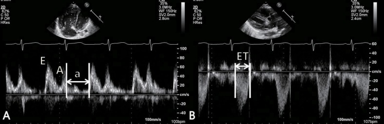
Despite developments in surgical techniques and other interventions, right ventricular (RV) failure remains an important clinical problem in several congenital heart diseases (CHD). RV function is one of the most important predictors of mortality and morbidity in patients with CHD. RV failure is a progressive disorder that begins with myocardial injury or stress, neurohormonal activation, cytokine activation, altered gene expression, and ventricular remodeling. Pressure-overload RV failure caused by RV outflow tract obstruction after total correction of tetralogy of Fallot, pulmonary stenosis, atrial switch operation for transposition of the great arteries, congenitally corrected transposition of the great arteries, and systemic RV failure after the Fontan operation. Volume-overload RV failure may be caused by atrial septal defect, pulmonary regurgitation, or tricuspid regurgitation. Although the measurement of RV function is difficult because of many reasons, the right ventricle can be evaluated using both imaging and functional modalities. In clinical practice, echocardiography is the primary mode for the evaluation of RV structure and function. Cardiac magnetic resonance imaging is increasingly used for evaluating RV structure and function. A comprehensive evaluation of RV function may lead to early and optimal management of RV failure in patients with CHD.
Keywords: Congenital heart disease; Right ventricle; Right-side heart failure.
Conflict of interest statement
No potential conflict of interest relevant to this article was reported.
Figures


References
-
- Voelkel NF, Quaife RA, Leinwand LA, Barst RJ, McGoon MD, Meldrum DR, et al. Right ventricular function and failure: report of a National Heart, Lung, and Blood Institute working group on cellular and molecular mechanisms of right heart failure. Circulation. 2006;114:1883–1891. - PubMed
-
- Haddad F, Hunt SA, Rosenthal DN, Murphy DJ. Right ventricular function in cardiovascular disease, part I: anatomy, physiology, aging, and functional assessment of the right ventricle. Circulation. 2008;117:1436–1448. - PubMed
-
- Haddad F, Couture P, Tousignant C, Denault AY. The right ventricle in cardiac surgery, a perioperative perspective: I. Anatomy, physiology, and assessment. Anesth Analg. 2009;108:407–421. - PubMed
LinkOut - more resources
Full Text Sources
Other Literature Sources