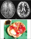
Giant cavernous hemangiomas of the brain
- PMID: 23559993
- PMCID: PMC3613648
- DOI: 10.4103/1793-5482.106660
Giant cavernous hemangiomas of the brain
Abstract
Cavernous angiomas or hemangiomas or cavernomas are benign, vascular malformations of the central nervous system and classified as occult vascular brain lesions, usually present in adulthood. With the advent of computed tomography and magnetic resonance imaging, these lesions are increasingly recognized in children. We report two cases of pediatric brain cavernomas where the children presented with recurrent seizures. Imaging findings were suggestive of giant cavernous hemangioma. The lesions were excised completely and the patients recovered well without deficits with good control of seizures.
Keywords: Cavernous hemangioma; epilepsy; giant; magnetic resonance imaging.
Conflict of interest statement
Figures


Similar articles
-
Paediatric giant cavernomas: report of three cases with a review of the literature.Childs Nerv Syst. 2021 Dec;37(12):3835-3845. doi: 10.1007/s00381-021-05286-6. Epub 2021 Jul 11. Childs Nerv Syst. 2021. PMID: 34247276 Review.
-
Intracranial extra-axial cavernous (HEM) angiomas: tumors or vascular malformations?J Neuroradiol. 2002 Jun;29(2):91-104. J Neuroradiol. 2002. PMID: 12297731 Review.
-
[Giant cavernous angioma: report of two cases].Arq Neuropsiquiatr. 2002 Jun;60(2-B):481-6. Arq Neuropsiquiatr. 2002. PMID: 12131955 Review. Portuguese.
-
Association between trauma and acute hemorrhage of cavernous malformations in children: report of 3 cases.J Neurosurg Pediatr. 2016 Sep;18(3):263-8. doi: 10.3171/2016.3.PEDS15517. Epub 2016 May 6. J Neurosurg Pediatr. 2016. PMID: 27153379
-
Cerebral cavernous angiomas in critical areas. Reports of three cases in children.J Neurosurg Sci. 1997 Dec;41(4):353-7. J Neurosurg Sci. 1997. PMID: 9555643
Cited by
-
Paediatric giant cavernomas: report of three cases with a review of the literature.Childs Nerv Syst. 2021 Dec;37(12):3835-3845. doi: 10.1007/s00381-021-05286-6. Epub 2021 Jul 11. Childs Nerv Syst. 2021. PMID: 34247276 Review.
-
Surgical treatment of cavernous malformation-related epilepsy in children: case series, systematic review, and meta-analysis.Neurosurg Rev. 2024 May 31;47(1):251. doi: 10.1007/s10143-024-02491-0. Neurosurg Rev. 2024. PMID: 38819574
-
A giant frontal cavernous malformation with review of literature.J Neurosci Rural Pract. 2016 Apr-Jun;7(2):279-82. doi: 10.4103/0976-3147.178666. J Neurosci Rural Pract. 2016. PMID: 27114662 Free PMC article.
-
Rare case of giant pediatric cavernous angioma of the temporal lobe: A case report and review of the literature.Surg Neurol Int. 2020 Jan 10;11:7. doi: 10.25259/SNI_468_2019. eCollection 2020. Surg Neurol Int. 2020. PMID: 31966926 Free PMC article.
-
Hyper-vascular giant cavernous malformation in a child: a case report and review.Childs Nerv Syst. 2017 Feb;33(2):375-379. doi: 10.1007/s00381-016-3234-8. Epub 2016 Sep 1. Childs Nerv Syst. 2017. PMID: 27585994 Review.
References
-
- de Andrade GC, Prandini MN, Braga FM. [Giant cavernous angioma: report of two cases] Arq Neuropsiquiatr. 2002;60:481–6. - PubMed
-
- Buckingham MJ, Crone KR, Ball WS, Berger TS. Management of cerebral cavernous angiomas in children presenting with seizures. Childs Nerv Syst. 1989;5:347–9. - PubMed
-
- van Lindert EJ, Tan TC, Grotenhuis JA, Wesseling P. Giant cavernous hemangiomas: Report of three cases. Neurosurg Rev. 2007;30:83–92. - PubMed
-
- Seifert V, Trost HA, Dietz H. Cavernous angiomas of the supratentorial compartment. Zentralbl Neurochir. 1989;50:89–92. - PubMed
-
- Giulioni M, Acciarri N, Padovani R, Frank F, Galassi E, Gaist G. Surgical management of cavernous angiomas in children. Surg Neurol. 1994;42:194–9. - PubMed

