

Chondromyxoid fibroma of the temporal bone: A rare entity
- PMID: 23560012
- PMCID: PMC3611914
- DOI: 10.4103/1817-1745.106483
Chondromyxoid fibroma of the temporal bone: A rare entity
Abstract
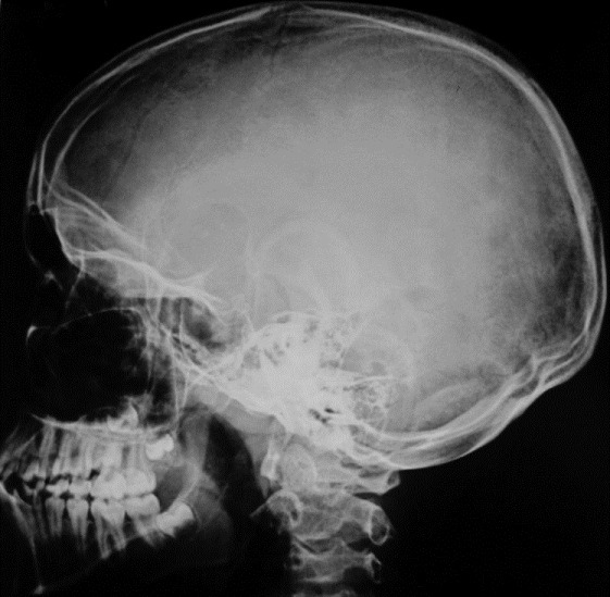
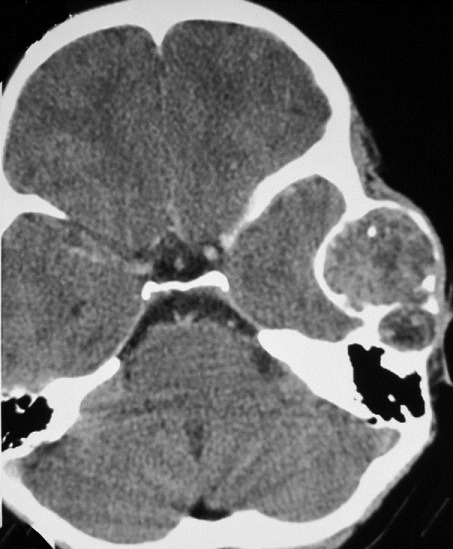
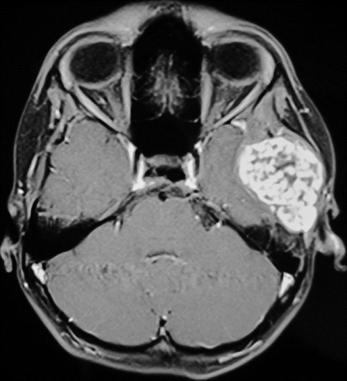
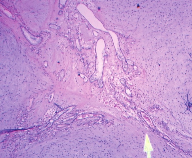
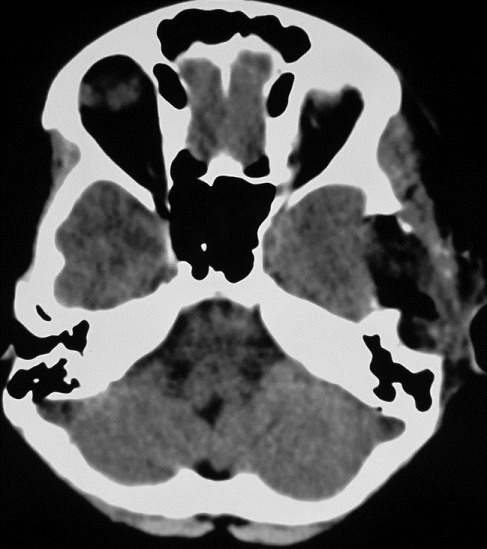
Chondromyxoid fibroma (CMF) is the least common benign tumor of the cartilaginous origin. It is very unusual to find these tumors in the skull bones. We report one such case involving the temporal bone. Till date, only nine such cases including this patient, involving the temporal bone have been reported to the best of our knowledge. Grant Medical College and Sir J.J Group of Hospitals, Byculla, Mumbai, Maharashtra, India. A 12-year-old female patient presented with a history of headache associated with left earache of 1 month duration. This was followed by swelling over the left preauricular region 15 days later. Imaging was suggestive of an expansile lesion involving the squamous part of the left temporal bone with calcifications suggestive of a benign chondroid lesion. The patient was operated upon with left temporal incision and complete excision of the lesion. The patient had relief from headache, earache and swelling, with no evidence of new neurological deficit in the post-operative period. CMF of the skull bone is an extremely rare tumor. Differential diagnosis should be kept in mind, especially in cases of calcified lesions and includes chordoma, chondroid chondroma, and low-grade myxoid chondrosarcoma. En-bloc complete excision should be the aim to achieve cure.
Keywords: Chondromyxoid; fibroma; temporal.
Conflict of interest statement
Figures
Similar articles
-
Chondromyxoid Fibroma of the Mastoid: A Rare Entity with Comprehensive Literature Review.J Int Adv Otol. 2020 Apr;16(1):117-122. doi: 10.5152/iao.2019.6911. J Int Adv Otol. 2020. PMID: 32209521 Free PMC article. Review.
-
Chondromyxoid Fibroma of Distal Phalanx of the Great Toe: A Rare Clinical Entity.Cureus. 2020 Feb 28;12(2):e7133. doi: 10.7759/cureus.7133. Cureus. 2020. PMID: 32257678 Free PMC article.
-
Chondromyxoid Fibroma: A Rare Case Report and Review of Literature.Cureus. 2016 Sep 23;8(9):e803. doi: 10.7759/cureus.803. Cureus. 2016. PMID: 27833828 Free PMC article.
-
Chondromyxoid fibroma of the temporal bone: case report and review of the literature.Ann Otol Rhinol Laryngol. 2007 Dec;116(12):922-7. doi: 10.1177/000348940711601209. Ann Otol Rhinol Laryngol. 2007. PMID: 18217512
-
Chondromyxoid fibroma of sphenoid sinus with unusual calcifications: case report with literature review.Head Neck Pathol. 2009 Jun;3(2):169-73. doi: 10.1007/s12105-009-0121-6. Epub 2009 Jun 10. Head Neck Pathol. 2009. PMID: 19644549 Free PMC article. Review.
Cited by
-
Chondromyxoid fibroma of the temporal bone: A case report and review of the literature.World J Clin Cases. 2018 Dec 26;6(16):1210-1216. doi: 10.12998/wjcc.v6.i16.1210. World J Clin Cases. 2018. PMID: 30613685 Free PMC article.
-
Chondromyxoid fibroma of the temporal bone: A rare case report.Medicine (Baltimore). 2020 Mar;99(11):e19487. doi: 10.1097/MD.0000000000019487. Medicine (Baltimore). 2020. PMID: 32176085 Free PMC article. Review.
-
Evaluation of Skull Base Tumors with Dynamic TurboFLASH MRI during Gadolinium Injection.J Neurol Surg B Skull Base. 2020 Apr;81(2):172-179. doi: 10.1055/s-0039-1681039. Epub 2019 Mar 15. J Neurol Surg B Skull Base. 2020. PMID: 32206536 Free PMC article.
-
Chondromyxoid Fibroma of the Mastoid: A Rare Entity with Comprehensive Literature Review.J Int Adv Otol. 2020 Apr;16(1):117-122. doi: 10.5152/iao.2019.6911. J Int Adv Otol. 2020. PMID: 32209521 Free PMC article. Review.
References
-
- Jaffe HL, Lichtenstein L. Chondromyxoid fibroma of bone; a distinctive benign tumor likely to be mistaken especially for chondrosarcoma. Arch Pathol (Chic) 1948;45:541–51. - PubMed
-
- Tarhan NC, Yologlu Z, Tutar NU, Coskun M, Agildere AM, Arikan U. Chondromyxoid fibroma of the temporal bone: CT and MRI findings. Eur Radiol. 2000;10:1678–80. - PubMed
-
- Wu CT, Inwards CY, O’Laughlin S, Rock MG, Beabout JW, Unni KK. Chondromyxoid fibroma of bone: A clinicopathologic review of 278 cases. Hum Pathol. 1998;29:438–46. - PubMed
-
- Haberal AN, Bulezuk B, Coskun M, Altinors N, Demirhan B. Unusual presentation of a chondromyxoid fibroma of the temporal bone. Turk J Med Sci. 2001;31:91–3.
-
- Unni KK. Dahlin's Bone Tumors General Aspects and Data on 11087 Cases. 5th ed. Philadelphia: Lippincott-Raven; 1996. Chondromyxoid fibroma; pp. 59–69.
Publication types
LinkOut - more resources
Full Text Sources
Research Materials
