Kinesin 5B (KIF5B) is required for progression through female meiosis and proper chromosomal segregation in mitotic cells
- PMID: 23560038
- PMCID: PMC3613343
- DOI: 10.1371/journal.pone.0058585
Kinesin 5B (KIF5B) is required for progression through female meiosis and proper chromosomal segregation in mitotic cells
Abstract
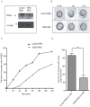
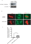
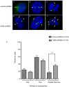
The fidelity of chromosomal segregation during cell division is important to maintain chromosomal stability in order to prevent cancer and birth defects. Although several spindle-associated molecular motors have been shown to be essential for cell division, only a few chromosome arm-associated motors have been described. Here, we investigated the role of Kinesin 5b (Kif5b) during female mouse meiotic cell development and mitotic cell division. RNA interference (RNAi)-mediated silencing of Kif5b in mouse oocytes induced significant delay in germinal vesicle breakdown (GVBD) and failure in extrusion of the first polar body (PBE). In mitotic cells, knockdown of Kif5b leads to centrosome amplification and a chromosomal segregation defect. These data suggest that KIF5B is critical in suppressing chromosomal instability at the early stages of female meiotic cell development and mitotic cell division.
Conflict of interest statement
Figures


Similar articles
-
CDCA8 regulates meiotic spindle assembly and chromosome segregation during human oocyte meiosis.Gene. 2020 May 30;741:144495. doi: 10.1016/j.gene.2020.144495. Epub 2020 Feb 20. Gene. 2020. PMID: 32088244
-
HURP permits MTOC sorting for robust meiotic spindle bipolarity, similar to extra centrosome clustering in cancer cells.J Cell Biol. 2010 Dec 27;191(7):1251-60. doi: 10.1083/jcb.201005065. Epub 2010 Dec 20. J Cell Biol. 2010. PMID: 21173113 Free PMC article.
-
Shifting meiotic to mitotic spindle assembly in oocytes disrupts chromosome alignment.EMBO Rep. 2018 Feb;19(2):368-381. doi: 10.15252/embr.201745225. Epub 2018 Jan 12. EMBO Rep. 2018. PMID: 29330318 Free PMC article.
-
Mitotic spindle multipolarity without centrosome amplification.Nat Cell Biol. 2014 May;16(5):386-94. doi: 10.1038/ncb2958. Nat Cell Biol. 2014. PMID: 24914434 Review.
-
Motoring through: the role of kinesin superfamily proteins in female meiosis.Hum Reprod Update. 2017 Jul 1;23(4):409-420. doi: 10.1093/humupd/dmx010. Hum Reprod Update. 2017. PMID: 28431155 Review.
Cited by
-
Role of Antizyme Inhibitor Proteins in Cancers and Beyond.Onco Targets Ther. 2021 Jan 25;14:667-682. doi: 10.2147/OTT.S281157. eCollection 2021. Onco Targets Ther. 2021. PMID: 33531815 Free PMC article. Review.
-
HURP localization in metaphase is the result of a multi-step process requiring its phosphorylation at Ser627 residue.Front Cell Dev Biol. 2023 Jul 5;11:981425. doi: 10.3389/fcell.2023.981425. eCollection 2023. Front Cell Dev Biol. 2023. PMID: 37484914 Free PMC article.
-
The cohesin-associated protein Pds5A governs the meiotic spindle assembly via deubiquitination of Kif5B in oocytes.Sci Adv. 2025 Apr 11;11(15):eadt6159. doi: 10.1126/sciadv.adt6159. Epub 2025 Apr 11. Sci Adv. 2025. PMID: 40215310 Free PMC article.
-
ZP3 is Required for Germinal Vesicle Breakdown in Mouse Oocyte Meiosis.Sci Rep. 2017 Feb 1;7:41272. doi: 10.1038/srep41272. Sci Rep. 2017. PMID: 28145526 Free PMC article.
-
Centrifugal Displacement of Nuclei Reveals Multiple LINC Complex Mechanisms for Homeostatic Nuclear Positioning.Curr Biol. 2017 Oct 23;27(20):3097-3110.e5. doi: 10.1016/j.cub.2017.08.073. Epub 2017 Oct 5. Curr Biol. 2017. PMID: 28988861 Free PMC article.
References
-
- Rieder CL, Maiato H (2004) Stuck in division or passing through: what happens when cells cannot satisfy the spindle assembly checkpoint. Dev Cell 7: 637–651. - PubMed
-
- Musacchio A, Salmon ED (2007) The spindle – assembly checkpoint in space an time. Nat Rev Mol Cell Biol 8: 379–393. - PubMed
-
- Vallente RU, Cheng EY, Hassold TJ (2006) The synaptonemal complex and meiotic recombination in humans: new approaches to old questions. Chromosoma 115: 241. - PubMed
-
- Szollosi D, Calarco P, Donahue RP (1972) Absence of centrioles in the first and second meiotic spindles of mouse oocytes. J Cell Sci 11: 521–541. - PubMed
-
- Marshall WF (2002) Polar wind left flapping in the breeze? Trends Cell Biol 12: 9. - PubMed
Publication types
MeSH terms
Substances
Grants and funding
LinkOut - more resources
Full Text Sources
Other Literature Sources
Molecular Biology Databases
Miscellaneous

