

Statistical analysis of sleep spindle occurrences
- PMID: 23560045
- PMCID: PMC3613364
- DOI: 10.1371/journal.pone.0059318
Statistical analysis of sleep spindle occurrences
Abstract
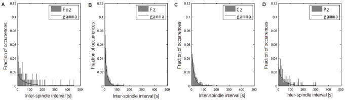
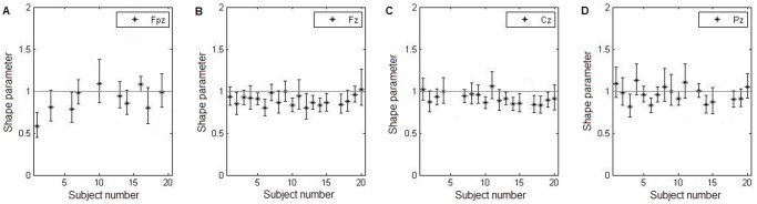
Spindles - a hallmark of stage II sleep - are a transient oscillatory phenomenon in the EEG believed to reflect thalamocortical activity contributing to unresponsiveness during sleep. Currently spindles are often classified into two classes: fast spindles, with a frequency of around 14 Hz, occurring in the centro-parietal region; and slow spindles, with a frequency of around 12 Hz, prevalent in the frontal region. Here we aim to establish whether the spindle generation process also exhibits spatial heterogeneity. Electroencephalographic recordings from 20 subjects were automatically scanned to detect spindles and the time occurrences of spindles were used for statistical analysis. Gamma distribution parameters were fit to each inter-spindle interval distribution, and a modified Wald-Wolfowitz lag-1 correlation test was applied. Results indicate that not all spindles are generated by the same statistical process, but this dissociation is not spindle-type specific. Although this dissociation is not topographically specific, a single generator for all spindle types appears unlikely.
Conflict of interest statement
Figures


Similar articles
-
Spatiotemporal changes of slow wave activities before and after 14 Hz/12 Hz sleep spindles during stage 2 sleep.Psychiatry Clin Neurosci. 2001 Jun;55(3):183-4. doi: 10.1046/j.1440-1819.2001.00817.x. Psychiatry Clin Neurosci. 2001. PMID: 11422833
-
[Developmental characteristics of frontal spindle and centro-parietal spindle].No To Hattatsu. 1996 Sep;28(5):409-17. No To Hattatsu. 1996. PMID: 8831244 Japanese.
-
Activation of fast sleep spindles at the premotor cortex and parietal areas contributes to motor learning: a study using sLORETA.Clin Neurophysiol. 2009 May;120(5):878-86. doi: 10.1016/j.clinph.2009.03.006. Epub 2009 Apr 18. Clin Neurophysiol. 2009. PMID: 19376746
-
Sleep spindles: an overview.Sleep Med Rev. 2003 Oct;7(5):423-40. doi: 10.1053/smrv.2002.0252. Sleep Med Rev. 2003. PMID: 14573378 Review.
-
Sleep spindles as neurophysiological biomarkers of schizophrenia.Eur J Neurosci. 2024 Apr;59(8):1907-1917. doi: 10.1111/ejn.16178. Epub 2023 Oct 26. Eur J Neurosci. 2024. PMID: 37885306 Review.
Cited by
-
Sleep Spindles as an Electrographic Element: Description and Automatic Detection Methods.Neural Plast. 2016;2016:6783812. doi: 10.1155/2016/6783812. Epub 2016 Jul 11. Neural Plast. 2016. PMID: 27478649 Free PMC article. Review.
-
Circadian preference towards morningness is associated with lower slow sleep spindle amplitude and intensity in adolescents.Sci Rep. 2017 Nov 6;7(1):14619. doi: 10.1038/s41598-017-13846-7. Sci Rep. 2017. PMID: 29097698 Free PMC article.
-
Spindle oscillations in communicating axons within a reconstituted hippocampal formation are strongest in CA3 without thalamus.Sci Rep. 2024 Apr 10;14(1):8384. doi: 10.1038/s41598-024-58002-0. Sci Rep. 2024. PMID: 38600114 Free PMC article.
-
Thalamocortical and intracortical laminar connectivity determines sleep spindle properties.PLoS Comput Biol. 2018 Jun 27;14(6):e1006171. doi: 10.1371/journal.pcbi.1006171. eCollection 2018 Jun. PLoS Comput Biol. 2018. PMID: 29949575 Free PMC article.
References
-
- Rechtschaffen A, Kales A (1968) A manual of standardized terminology, techniques and scoring system for sleep stages in human subjects, National Institutes of Health Publications. Washington DC: US Government Printing Office.
-
- Żygierewicz J, Blinowska KJ, Durka PJ, Szelenberger W, Niemcewicz Sz, et al. (1999) High resolution study of sleep spindles. Clin Neurophysiol 110: 2136–2147. - PubMed
-
- De Gennaro L, Ferrara M (2003) Sleep spindles: an overview. Sleep Medicine Rev 7(5): 423–440. - PubMed
-
- Sejnowski TJ, Destexhe A (2000) Why do we sleep? Brain Res 886: 208–223. - PubMed
Publication types
MeSH terms
Grants and funding
LinkOut - more resources
Full Text Sources
Other Literature Sources
