. 2013;8(4):e60261.
doi: 10.1371/journal.pone.0060261. Epub 2013 Apr 1.

New components of Drosophila leg development identified through genome wide association studies

Affiliations

New components of Drosophila leg development identified through genome wide association studies

Nathaniel Grubbs et al. PLoS One. 2013.

Abstract

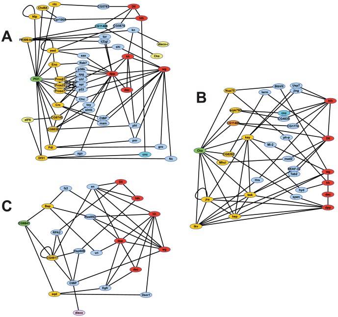
The adult Drosophila melanogaster body develops from imaginal discs, groups of cells set-aside during embryogenesis and expanded in number during larval stages. Specification and development of Drosophila imaginal discs have been studied for many years as models of morphogenesis. These studies are often based on mutations with large developmental effects, mutations that are often lethal in embryos when homozygous. Such forward genetic screens can be limited by factors such as early lethality and genetic redundancy. To identify additional genes and genetic pathways involved in leg imaginal disc development, we employed a Genome Wide Association Study utilizing the natural genetic variation in leg proportionality found in the Drosophila Genetic Reference Panel fly lines. In addition to identifying genes already known to be involved in leg development, we identified several genes involved in pathways that had not previously been linked with leg development. Several of the genes appear to be involved in signaling activities, while others have no known roles at this time. Many of these uncharacterized genes are conserved in mammals, so we can now begin to place these genes into developmental contexts. Interestingly, we identified five genes which, when their function is reduced by RNAi, cause an antenna-to-leg transformation. Our results demonstrate the utility of this approach, integrating the tools of quantitative and molecular genetics to study developmental processes, and provide new insights into the pathways and networks involved in Drosophila leg development.


Conflict of interest statement

Competing Interests: The authors have declared that no competing interests exist.

Figures

Figure 1
Figure 1. Model for larval leg disc patterning, proportionality and adult leg structure.
A) Leg imaginal disc patterning begins (left) when En, present in the posterior (yellow) portion of the disc, activates hh expression. The Hh ligand diffuses into the anterior portion of the disc. There it activates the expression of dpp (purple) dorsally and wg (green) ventrally. Dpp and Wg, in turn, diffuse throughout the dorsal and ventral portions, respectively, of the disc, creating a gradient of their mutual presence. This gradient is responsible for the pattern of expression of transcription factors (middle) that establish the proximal-distal axis of the leg (right). In the center of the disc, mutual Dpp and Wg is highest, activating the expression of Dll, which is responsible for patterning the most distal structures of the leg, including the terminal structures (claw and pulvillus). Reduced mutual Dpp and Wg results in the activation of dac, responsible for the patterning of the middle portions of the leg. Where there is almost no mutual Dpp or Wg, Hth and Exd are active, patterning the proximal leg portions and the junction with the rest of the body. B–C illustrate how proportions might change without affecting over-all length. B) Enhanced Hh signal (dark yellow) could result in an expansion of Wg and Dpp, causing a broader domain of Dll expression, at the expense of dac (middle). This could, in turn, cause the tarsal segments to make up a larger portion of the leg without changing leg length (right). C) Similarly, the proportion of the femur could be expanded (right) if dac expression were expanded (middle). This might result from a decrease in expression or distribution of Dpp and Wg (left), which could, in turn, be caused by reduced Hh signal (light yellow).
Figure 2
Figure 2. Distribution of candidate gene mRNA in imaginal discs.
Wild-type third instar imaginal discs stained to detect mRNAs from several of our candidate genes. Examples with several different mRNA distribution patterns are shown. There are several that are of note, the cross-like pattern of boi in the wing disc that borders wg and dpp expression, the narrow tarsal staining of eIF6. We also note that disco-r is expressed in the ventral edge of the wing which had not been reported previously.
Figure 3
Figure 3. Dll-Gal4 driving RNAi phenocopies in legs.
Second thoracic legs from female flies with Dll-Gal4 driving UAS-RNAi insertions. All flies were grown at room temperature. A) Wild-type. B) RNAi of CSN1b resulted in reduced leg size, but did not alter shape. C) RNAi of PI31 reduced length of tarsal segments, with occasional loss of a single distal segment. D) RNAi of eIF6 caused deletions and fusions of tarsal segments. E) RNAi of CG6841 deleted most of the tarsal segments and terminal structures. F) RNAi of Cka was similar to that of CG6841, but more severe. Abbreviations: fe, femur; ti, tibia; ta, tarsal segments; cl, claw.
Figure 4
Figure 4. Effect in antennae of Dll-Gal4 driving RNAi.
Heads from flies with both Dll-Gal4 and UAS-RNAi insertions that disrupted antennal development. Arrows with numbers indicate the antennal segments. A) Wild-type. B) RNAi of CSN1b resulted in loss of arista, and appearance of sclerotized structure C). RNAi of PI31 resulted in loss of arista while the remaining structures appear normal. D) RNAi of CG6841 resulted in loss of arista, and drastic changes to the appearance of antennal segment 3, and some reduction of A2. E) RNAi of Cka resulted in loss of all but proximal structures. F) RNAi of CG9129 is characteristic of the five lines that resulted in transformation of antennae toward leg identity beginning in distal A2, though slight differences were noted between the five lines that caused these transformations.
Figure 5
Figure 5. DroID analyses identifying connections between candidates and canonical leg development genes.
Genes in orange connect directly to the candidate (Green), and may be linked directly or through genes in blue to the canonical appendage cascade genes (red). Yellow identifies those of our candidate genes that are linked to the test gene. A) Analysis of PI31. Links shared with Cka are shown in dark blue. B) Analysis of Cka. Links shared with PI31 are shown in dark orange and dark blue. C) Analysis of CG6841, including a link to disco in pink.

References

    1. Kojima T (2004) The mechanism of Drosophila leg development along the proximodistal axis. Dev Growth Differ 46: 115–129. - PubMed
    1. Morata G (2001) How Drosophila appendages develop. Nat Rev Mol Cell Biol 2: 89–97. - PubMed
    1. Pueyo JI, Couso JP (2005) Parallels between the proximal-distal development of vertebrate and arthropod appendages: homology without an ancestor? Curr Opin Genet Dev 15: 439–446. - PubMed
    1. Estella C, Voutev R, Mann RS (2012) A dynamic network of morphogens and transcription factors patterns the fly leg. Curr Top Dev Biol 98: 173–198. - PMC - PubMed
    1. Panganiban G (2000) Distal-less function during Drosophila appendage and sense organ development. Dev Dyn 218: 554–562. - PubMed

Publication types

MeSH terms

LinkOut - more resources