. 2013;8(4):e60335.
doi: 10.1371/journal.pone.0060335. Epub 2013 Apr 1.

Transforming growth factor β1 induces the expression of collagen type I by DNA methylation in cardiac fibroblasts

Affiliations

Transforming growth factor β1 induces the expression of collagen type I by DNA methylation in cardiac fibroblasts

Xiaodong Pan et al. PLoS One. 2013.

Abstract

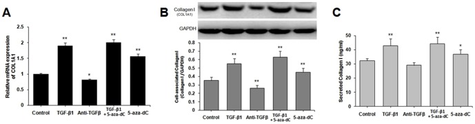
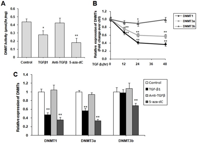
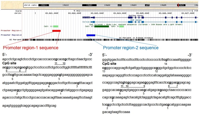
Transforming growth factor-beta (TGF-β), a key mediator of cardiac fibroblast activation, has a major influence on collagen type I production. However, the epigenetic mechanisms by which TGF-β induces collagen type I alpha 1 (COL1A1) expression are not fully understood. This study was designed to examine whether or not DNA methylation is involved in TGF-β-induced COL1A1 expression in cardiac fibroblasts. Cells isolated from neonatal Sprague-Dawley rats were cultured and stimulated with TGF-β1. The mRNA levels of COL1A1 and DNA methyltransferases (DNMTs) were determined via quantitative polymerase chain reaction and the protein levels of collagen type I were determined via Western blot as well as enzyme-linked immunosorbent assay. The quantitative methylation of the COL1A1 promoter region was analyzed using the MassARRAY platform of Sequenom. Results showed that TGF-β1 upregulated the mRNA expression of COL1A1 and induced the synthesis of cell-associated and secreted collagen type I in cardiac fibroblasts. DNMT1 and DNMT3a expressions were significantly downregulated and the global DNMT activity was inhibited when treated with 10 ng/mL of TGF-β1 for 48 h. TGF-β1 treatment resulted in a significant reduction of the DNA methylation percentage across multiple CpG sites in the rat COL1A1 promoter. Thus, TGF-β1 can induce collagen type I expression through the inhibition of DNMT1 and DNMT3a expressions as well as global DNMT activity, thereby resulting in DNA demethylation of the COL1A1 promoter. These findings suggested that the DNMT-mediated DNA methylation is an important mechanism in regulating the TGF-β1-induced COL1A1 gene expression.


Conflict of interest statement

Competing Interests: The authors have declared that no competing interests exist.

Figures

Figure 1
Figure 1. TGF-β1 and DNMT inhibitor induced the expression of collagen type I (COL1A1).
Untreated cardiac fibroblasts (CFs) were cultured until near confluence was reached, and then starved for 12 h in serum-free DMEM. CFs were stimulated with 10 ng/mL of TGF-β1, 30 µg/mL of TGF-β-neutralizing antibody, 10 ng/mL TGF-β1+5 µM 5-aza-dC and 5 µM 5-aza-dC for 48 h. (A) Collagen type I (COL1A1) mRNA was determined via quantitative real-time PCR. (B) Cell-associated collagen type I was determined via Western blot. (C) Secreted collagen type I was determined via ELISA. Data are presented as mean ± SD (n = 3). *P<0.05, **P<0.01 (relative to the respective control).
Figure 2
Figure 2. TGF-β1 inhibited the expression of DNMTs in cardiac fibroblasts (CFs).
(A) CFs were starved for 12 h in serum-free DMEM, and then stimulated with 10 ng/mL TGF-β1, 30 µg/mL TGF-β-neutralizing antibody and 5 µM 5-aza-dC for 48 h. A DNMT activity assay kit was used to analyze the global DNMT activity. (B) CFs were treated with 10 ng/mL TGF-β1 for 0, 12, 24 and 48 h. The expression of three DNMTs were analyzed by quantitative real-time PCR. (C) CFs were treat as (A), qPCR was performed to quantify the relative mRNA levels of DNMT1, DNMT3a, and DNMT3b. Data were obtained from three independent experiments and expressed as mean ± SD (n = 3). *P <0.05, **P<0.01 (relative to the respective control).
Figure 3
Figure 3. Schematic diagram of rat COL1A1 promoter.
Two DNA fragments from rat distal and proximal promoters were amplified to analyze the methylation of the COL1A1 promoter. The fragments were labeled as promoter region-1 and promoter region-2. The location of promoter region-1 (–1682 bp to –1322 bp), promoter region-2 (–184 bp to +199 bp), and three CpG islands are indicated by a red bar, a blue bar, and three green bars, respectively. The start of exon 1 was considered as +1 of the sequence. PCR primers were designed based on the reverse complementary strands of these fragments. Promoter region-1 sequence represents 360 bp fragments and the CpG sites were numbered from 1 to 11 from the 3′-end to the 5′-end. Promoter region-2 sequence represents 384 bp fragments and the CpG sites were numbered from 1 to 14 from the 3′-end to the 5′-end. The numbers refer to the locations of the CpG sites. The underlined highlights correspond to the multiple CpG sites that were tested simultaneously.
Figure 4
Figure 4. Methylation levels of the CpG sites in the COL1A1 promoter.
The methylation levels of CpG sites in COL1A1 promoter regions from the control group (without treatment), TGF-β group (treated with 10 ng/mL TGF-β1 for 48 h) and 5-aza-dC group (treated with 5 µM 5-aza-dC for 48 h) were compared. Sequenom MassARRAY platform was used for the quantitative methylation analysis. The colors of each circle represent the methylation level of each corresponding CpG unit. Quantitative methylation analysis results are shown in a color scale: yellow (∼0% methylation), green (∼50% methylation), and dark blue (∼100% methylation). The white circles represent the missing data at a given CpG site. Mean methylation levels of CpG sites in (A) COL1A1 promoter region-1 and (B) COL1A1 promoter region-2. Data are expressed as mean ± SD∶ n control = 5 (Sample: C-1 to C-5); n TGF-β  = 6 (Sample: T4-1 to T4-6); n 5-aza-dC = 3(Sample: A-1 to A-3). *P <0.05, **P <0.01 (relative to the respective control).

References

    1. Brown RD, Ambler SK, Mitchell MD, Long CS (2005) The cardiac fibroblast: therapeutic target in myocardial remodeling and failure. Annu Rev Pharmacol Toxicol 45: 657–687. - PubMed
    1. Jugdutt BI (2003) Remodeling of the myocardium and potential targets in the collagen degradation and synthesis pathways. Curr Drug Targets Cardiovasc Haematol Disord 3: 1–30. - PubMed
    1. Porter KE, Turner NA (2009) Cardiac fibroblasts: at the heart of myocardial remodeling. Pharmacol Ther 123: 255–278. - PubMed
    1. Berk BC, Fujiwara K, Lehoux S (2007) ECM remodeling in hypertensive heart disease. J Clin Investig 117: 568–575. - PMC - PubMed
    1. Leask A, Abraham DJ (2004) TGF-beta signaling and the fibrotic response. FASEB J 18: 816–827. - PubMed

Publication types

MeSH terms

Substances