

Lymphoma B-cell responsiveness to CpG-DNA depends on the tumor microenvironment
- PMID: 23561041
- PMCID: PMC3623776
- DOI: 10.1186/1756-9966-32-18
Lymphoma B-cell responsiveness to CpG-DNA depends on the tumor microenvironment
Abstract
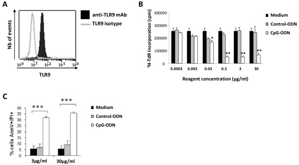
Background: Toll-like receptor (TLR) agonists have important properties that can be exploited for immunotherapy against tumors. Locally injected immunostimulatory oligodeoxynucleotides containing CpG motifs (CpG-ODNs), which are TLR9 agonists, have shown promise in cancer models. Several studies have demonstrated that these motifs have immunologic effects similar to those of bacterial DNA and can stimulate monocytes, macrophages, dendritic, and B cells, which then produce several proinflammatory cytokines. However, these CpG-ODNs appear to produce opposite effects on tumor B cells.
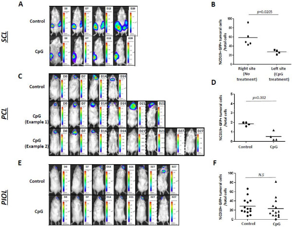
Methods: In this study, we investigated the direct effects of a murine class B CpG (1826) ODNs on lymphoma B cells in vitro and in vivo, using mouse models of non-Hodgkin B lymphomas developing in immunoprivileged sites, specifically the brain and the eye, and in subcutaneous sites.
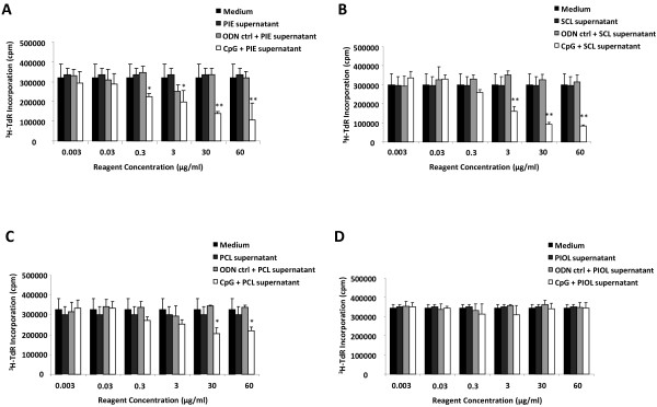
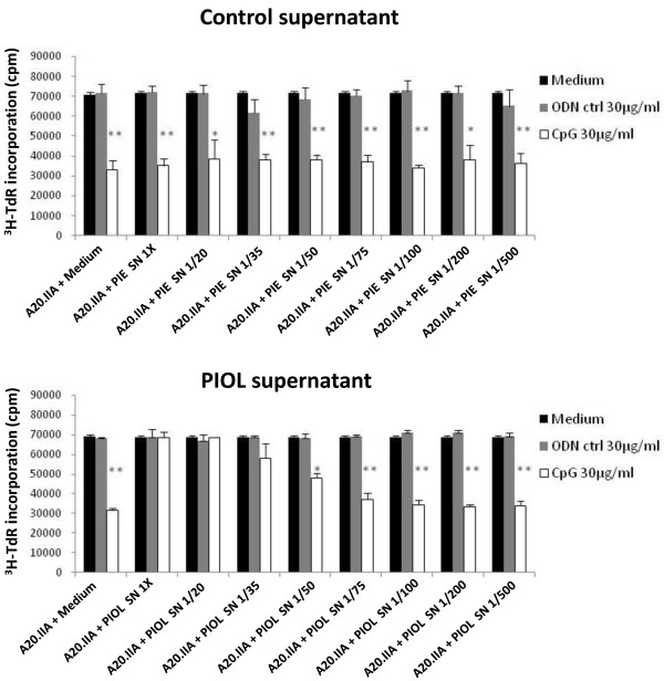
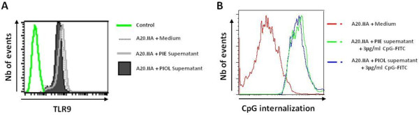
Results: In vitro, CpG-ODNs produced antiproliferative and proapoptotic effects on lymphoma B cells. In vivo, it had an antitumor effect when injected into tumors in murine models of subcutaneous lymphoma (SCL) and primary cerebral lymphoma (PCL). However, its intravitreal administration into a primary intraocular lymphoma (PIOL) mouse model did not produce an antitumor effect. In vitro experiments using supernatant from mouse PIOL samples demonstrated that the PIOL molecular microenvironment inhibits the antiproliferative effect of CpG-ODNs on lymphoma B-cells.
Conclusions: Responsiveness to CpG stimulation differs in subcutaneous, cerebral, and ocular tumors, according to the tumoral and molecular microenvironment, and this should be considered for further therapeutic approaches.
Figures
Similar articles
-
Toll-like receptor agonists induce apoptosis in mouse B-cell lymphoma cells by altering NF-κB activation.Cell Mol Immunol. 2013 Jul;10(4):360-72. doi: 10.1038/cmi.2013.14. Epub 2013 Jun 3. Cell Mol Immunol. 2013. PMID: 23727784 Free PMC article.
-
Lymphoma immunotherapy with CpG oligodeoxynucleotides requires TLR9 either in the host or in the tumor itself.J Immunol. 2007 Aug 15;179(4):2493-500. doi: 10.4049/jimmunol.179.4.2493. J Immunol. 2007. PMID: 17675511
-
B-cell lymphomas differ in their responsiveness to CpG oligodeoxynucleotides.Clin Cancer Res. 2005 Feb 15;11(4):1490-9. doi: 10.1158/1078-0432.CCR-04-1890. Clin Cancer Res. 2005. PMID: 15746051
-
Immunostimulatory Activities of CpG-Oligodeoxynucleotides in Teleosts: Toll-Like Receptors 9 and 21.Front Immunol. 2019 Feb 8;10:179. doi: 10.3389/fimmu.2019.00179. eCollection 2019. Front Immunol. 2019. PMID: 30800129 Free PMC article. Review.
-
CpG oligodeoxynucleotides as TLR9 agonists: therapeutic application in allergy and asthma.BioDrugs. 2010 Aug 1;24(4):225-35. doi: 10.2165/11536140-000000000-00000. BioDrugs. 2010. PMID: 20623989 Review.
Cited by
-
Tunable PhenoCycler imaging of the murine pre-clinical tumour microenvironments.Cell Biosci. 2024 Feb 4;14(1):19. doi: 10.1186/s13578-024-01199-4. Cell Biosci. 2024. PMID: 38311785 Free PMC article.
-
Enhanced systemic antilymphoma immune response by photothermal therapy with CpG deoxynucleotide-coated nanoparticles.Blood Adv. 2022 Aug 9;6(15):4581-4592. doi: 10.1182/bloodadvances.2022008040. Blood Adv. 2022. PMID: 35687489 Free PMC article.
-
Bioluminescence-Based Tumor Quantification Method for Monitoring Tumor Progression and Treatment Effects in Mouse Lymphoma Models.J Vis Exp. 2016 Jul 7;(113):53609. doi: 10.3791/53609. J Vis Exp. 2016. PMID: 27501019 Free PMC article.
-
Heterogeneity of Toll-like receptor 9 signaling in B cell malignancies and its potential therapeutic application.J Transl Med. 2017 Feb 27;15(1):51. doi: 10.1186/s12967-017-1152-5. J Transl Med. 2017. PMID: 28241765 Free PMC article. Review.
-
Differential expression of Toll-like receptor (TLR) and B cell receptor (BCR) signaling molecules in primary diffuse large B-cell lymphoma of the central nervous system.J Neurooncol. 2015 Jan;121(2):289-96. doi: 10.1007/s11060-014-1655-3. Epub 2014 Nov 13. J Neurooncol. 2015. PMID: 25391967
References
-
- Verthelyi D, Ishii KJ, Gursel M, Takeshita F, Klinman DM. Human peripheral blood cells differentially recognize and respond to two distinct CPG motifs. J Immunol. 2001;166(4):2372–2377. - PubMed
Publication types
MeSH terms
Substances
LinkOut - more resources
Full Text Sources
Other Literature Sources
Research Materials
Miscellaneous
