. 2013 Apr 2;104(7):1435-44.
doi: 10.1016/j.bpj.2013.01.054.

The association of polar residues in the DAP12 homodimer: TOXCAT and molecular dynamics simulation studies

Affiliations

The association of polar residues in the DAP12 homodimer: TOXCAT and molecular dynamics simulation studies

Peng Wei et al. Biophys J. .

Abstract

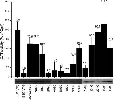
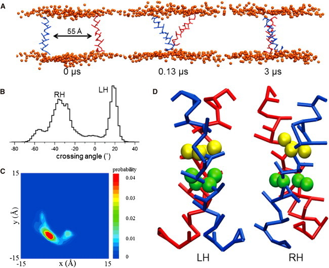
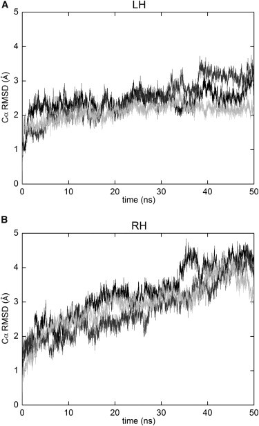
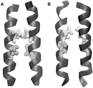
Dimerization of the transmembrane (TM) adaptor protein DAP12 plays a key role in mediating activation signals through TM-TM association with cell-surface receptors. Herein, we apply the TOXCAT assay and molecular dynamics simulation to analyze dynamics and dimerization of the TM helix of DAP12 in the membrane bilayer. In the TOXCAT assay, we performed site-specific mutagenesis of potential dimerization motifs in the DAP12 TM domain. Instead of the common GxxxG dimerization motif, mutating either of the polar residues Asp-50 and Thr-54 significantly decreased the TOXCAT signal for the dimerization of DAP12 TM domain. Furthermore, through the conformational difference between wild-type and mutant DAP12 TM homodimers, a combined coarse-grained and atomistic molecular dynamics simulation has identified both Asp-50 and Thr-54 at the dimerization interface. The experimental and computational results of the DAP12 TM dimer are in excellent agreement with the previously reported NMR structure obtained in detergent micelles. Such a combination of dynamics simulation and cell-based experiments can be applied to produce insights at the molecular level into the TM-TM association of many other transmembrane proteins.


Figures

Figure 1
Figure 1
DAP12-WT TM domain self-associated in the Escherichia coli cell membrane and the effect of site-specific mutagenesis in the GxxxG motif and polar residues D50 and T54 of the DAP12-WT TM domain on CAT activity. The enzymatic activity of CAT induced by self-association of the target TM domain and expressed as the percentage of that induced by the GpA-WT TM domain. The GpA-WT and GpA-G83I constructs were used as positive and negative controls, respectively. Black bars represent the CAT activity quantified from cleared lysates, and error bars represent the standard error for three measurements of each lysate. The lower panel shows the expression levels of chimeric ToxR-TM-MBP proteins probed by Western blot.
Figure 2
Figure 2
CG simulations to explore DAP12-WT helix homodimer. (A) The DAP12-WT helices were parallel and separated by ∼55 Å in the DPPC bilayer of the initial structure. During the 3 μs CG-WT simulations, they interact with each other and form a homodimer. (B) All the dimer trajectories of CG-WT simulations were converged and crossing angles were analyzed. The positive crossing angle corresponds to LH helix packing and the negative crossing angle corresponds to RH packing. (C) The spatial distribution of helix for the CG-WT simulations. The backbone particles were fitted to a reference structure. This diagram shows the contact probability density between the two TM helix backbone particles in the bilayer plane. The blue and red represents low probability and high probability at that point. The LH and RH dimer have only one maxima area in this distribution. (D) The LH and RH structures were selected to be representative of the modes in the crossing angle distributions. The backbone and side-chain particles of the D50 and T54 particles are in yellow and green.
Figure 3
Figure 3
The conformational stability of DAP12-WT helix homodimer in each atomistic simulation is analyzed based on the Cα RMSD from the initial structure as a function of time. The atomistic LH (A) and RH (B) representative initial helix dimer were conversed from the CG structures picked in the crossing angle distribution.
Figure 4
Figure 4
Comparison of the helix dimer interface of the NMR (2L34) structure (A) and a structure from the end of one of the AT-LH simulations (B). The D50 (white) are shown to pack against each other in the interface and the side chains of T54 (gray) stabilize the D50 around the interface. These key interactions are shown in both the NMR and the simulation structure.
Figure 5
Figure 5
Mutants of DAP12-WT helix homodimer crossing angle histograms. Helix crossing angle histograms. For each set of simulations, all the dimer frames were merged. The helix crossing angles were evaluated from these merged dimer trajectories. The positive crossing angle corresponds to LH helix packing and the negative crossing angle corresponds to RH packing. The WT is drawn in a solid line, and the mutants are drawn in a dashed line.
Figure 6
Figure 6
Spatial distributions of all the mutants of DAP12-WT helix. The backbone particles were fitted to a unique reference structure used in Fig. 2C. This diagram shows the contact probability density between the two TM helix backbone particles in the bilayer plane; white and black represent low and high probability.

Similar articles

Cited by

References

    1. Popot J.L., Engelman D.M. Membrane protein folding and oligomerization: the two-stage model. Biochemistry. 1990;29:4031–4037. - PubMed
    1. Melnyk R.A., Partridge A.W., Deber C.M. Transmembrane domain mediated self-assembly of major coat protein subunits from Ff bacteriophage. J. Mol. Biol. 2002;315:63–72. - PubMed
    1. Gaddie K.J., Kirley T.L. Conserved polar residues stabilize transmembrane domains and promote oligomerization in human nucleoside triphosphate diphosphohydrolase 3. Biochemistry. 2009;48:9437–9447. - PMC - PubMed
    1. Escher C., Cymer F., Schneider D. Two GxxxG-like motifs facilitate promiscuous interactions of the human ErbB transmembrane domains. J. Mol. Biol. 2009;389:10–16. - PubMed
    1. Treutlein H.R., Lemmon M.A., Brünger A.T. The glycophorin A transmembrane domain dimer: sequence-specific propensity for a right-handed supercoil of helices. Biochemistry. 1992;31:12726–12732. - PubMed

Publication types

MeSH terms

LinkOut - more resources