

Comparative studies of disordered proteins with similar sequences: application to Aβ40 and Aβ42
- PMID: 23561531
- PMCID: PMC3617440
- DOI: 10.1016/j.bpj.2013.02.023
Comparative studies of disordered proteins with similar sequences: application to Aβ40 and Aβ42
Abstract
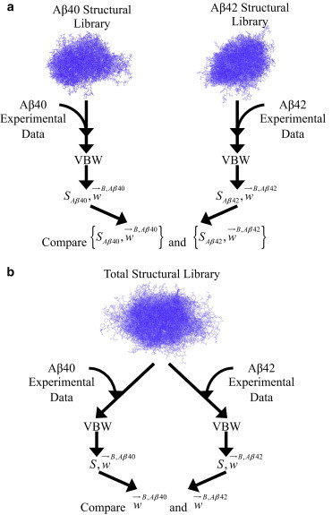
Quantitative comparisons of intrinsically disordered proteins (IDPs) with similar sequences, such as mutant forms of the same protein, may provide insights into IDP aggregation-a process that plays a role in several neurodegenerative disorders. Here we describe an approach for modeling IDPs with similar sequences that simplifies the comparison of the ensembles by utilizing a single library of structures. The relative population weights of the structures are estimated using a Bayesian formalism, which provides measures of uncertainty in the resulting ensembles. We applied this approach to the comparison of ensembles for Aβ40 and Aβ42. Bayesian hypothesis testing finds that although both Aβ species sample β-rich conformations in solution that may represent prefibrillar intermediates, the probability that Aβ42 samples these prefibrillar states is roughly an order of magnitude larger than the frequency in which Aβ40 samples such structures. Moreover, the structure of the soluble prefibrillar state in our ensembles is similar to the experimentally determined structure of Aβ that has been implicated as an intermediate in the aggregation pathway. Overall, our approach for comparative studies of IDPs with similar sequences provides a platform for future studies on the effect of mutations on the structure and function of disordered proteins.
Copyright © 2013 Biophysical Society. Published by Elsevier Inc. All rights reserved.
Figures


Similar articles
-
The structures of the E22Δ mutant-type amyloid-β alloforms and the impact of E22Δ mutation on the structures of the wild-type amyloid-β alloforms.ACS Chem Neurosci. 2013 Feb 20;4(2):310-20. doi: 10.1021/cn300149j. Epub 2012 Dec 18. ACS Chem Neurosci. 2013. PMID: 23421682 Free PMC article.
-
Homogeneous and heterogeneous tertiary structure ensembles of amyloid-β peptides.Biochemistry. 2011 Sep 6;50(35):7612-28. doi: 10.1021/bi200732x. Epub 2011 Aug 15. Biochemistry. 2011. PMID: 21797254 Free PMC article.
-
Differences in the free energies between the excited states of Aβ40 and Aβ42 monomers encode their aggregation propensities.Proc Natl Acad Sci U S A. 2020 Aug 18;117(33):19926-19937. doi: 10.1073/pnas.2002570117. Epub 2020 Jul 30. Proc Natl Acad Sci U S A. 2020. PMID: 32732434 Free PMC article.
-
Aβ42 and Aβ40: similarities and differences.J Pept Sci. 2015 Jul;21(7):522-9. doi: 10.1002/psc.2789. Epub 2015 May 28. J Pept Sci. 2015. PMID: 26018760 Review.
-
Role of the region 23-28 in Abeta fibril formation: insights from simulations of the monomers and dimers of Alzheimer's peptides Abeta40 and Abeta42.Curr Alzheimer Res. 2008 Jun;5(3):244-50. doi: 10.2174/156720508784533330. Curr Alzheimer Res. 2008. PMID: 18537541 Review.
Cited by
-
Seeding and Growth of β-Amyloid Aggregates upon Interaction with Neuronal Cell Membranes.Int J Mol Sci. 2020 Jul 16;21(14):5035. doi: 10.3390/ijms21145035. Int J Mol Sci. 2020. PMID: 32708806 Free PMC article.
-
Hamiltonian Switch Metropolis Monte Carlo Simulations for Improved Conformational Sampling of Intrinsically Disordered Regions Tethered to Ordered Domains of Proteins.J Chem Theory Comput. 2014 Aug 12;10(8):3550-3562. doi: 10.1021/ct5002297. Epub 2014 Jun 3. J Chem Theory Comput. 2014. PMID: 25136274 Free PMC article.
-
Spontaneous self-assembly of amyloid β (1-40) into dimers.Nanoscale Adv. 2019 Sep 17;1(10):3892-3899. doi: 10.1039/c9na00380k. eCollection 2019 Oct 9. Nanoscale Adv. 2019. PMID: 36132110 Free PMC article.
-
Comprehensive Structural and Thermodynamic Analysis of Prefibrillar WT α-Synuclein and Its G51D, E46K, and A53T Mutants by a Combination of Small-Angle X-ray Scattering and Variational Bayesian Weighting.J Chem Inf Model. 2020 Oct 26;60(10):5265-5281. doi: 10.1021/acs.jcim.0c00807. Epub 2020 Sep 17. J Chem Inf Model. 2020. PMID: 32866007 Free PMC article.
-
Digested disorder: Quarterly intrinsic disorder digest (April-May-June, 2013).Intrinsically Disord Proteins. 2013 Jan 1;1(1):e27454. doi: 10.4161/idp.27454. eCollection 2013 Jan-Dec. Intrinsically Disord Proteins. 2013. PMID: 28516028 Free PMC article. Review.
References
-
- Huang A., Stultz C.M. Finding order within disorder: elucidating the structure of proteins associated with neurodegenerative disease. Future Med. Chem. 2009;1:467–482. - PubMed
Publication types
MeSH terms
Substances
LinkOut - more resources
Full Text Sources
Other Literature Sources
