Review
. 2013 Apr 2;21(4):501-15.
doi: 10.1016/j.str.2013.02.024.

Apoptosome structure, assembly, and procaspase activation

Affiliations
Review

Apoptosome structure, assembly, and procaspase activation

Shujun Yuan et al. Structure. .

Abstract

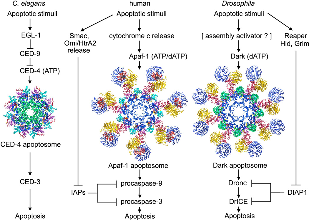
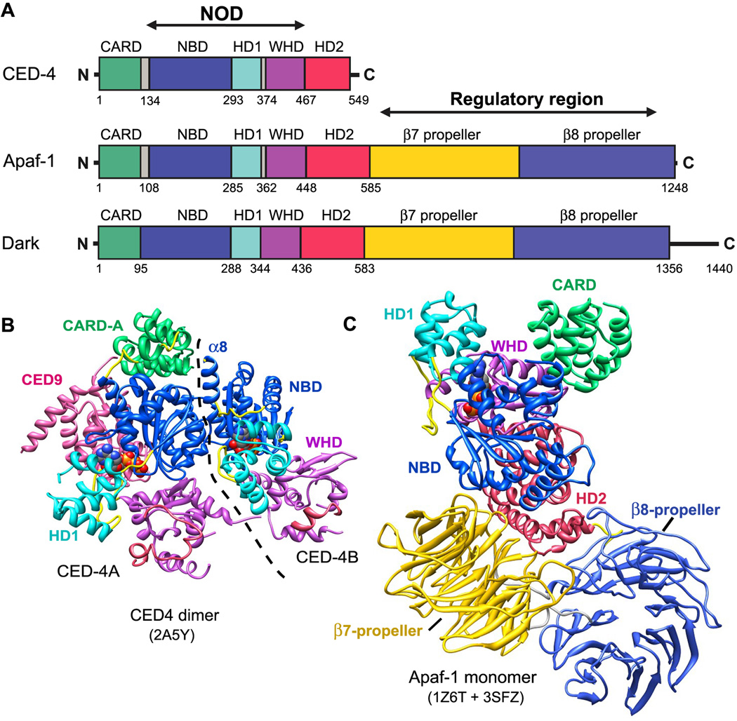
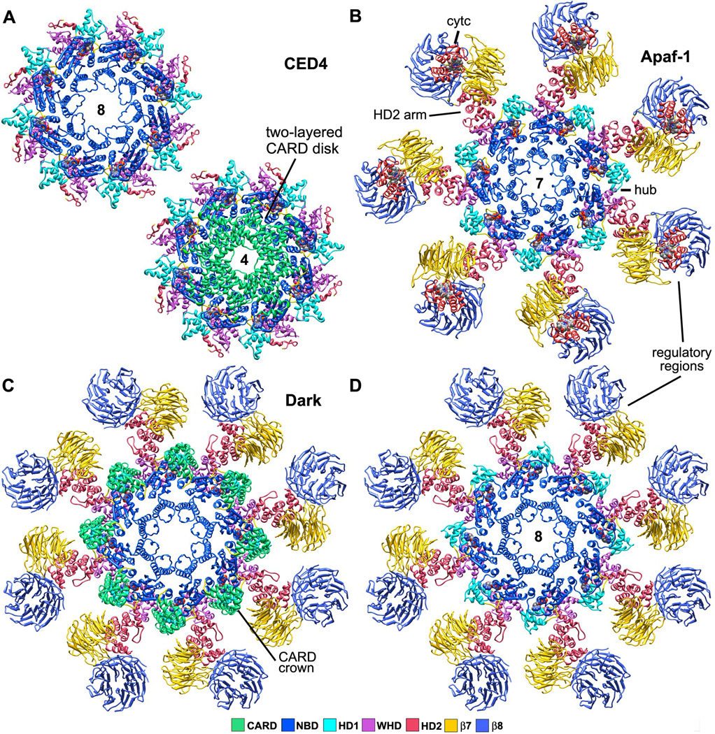
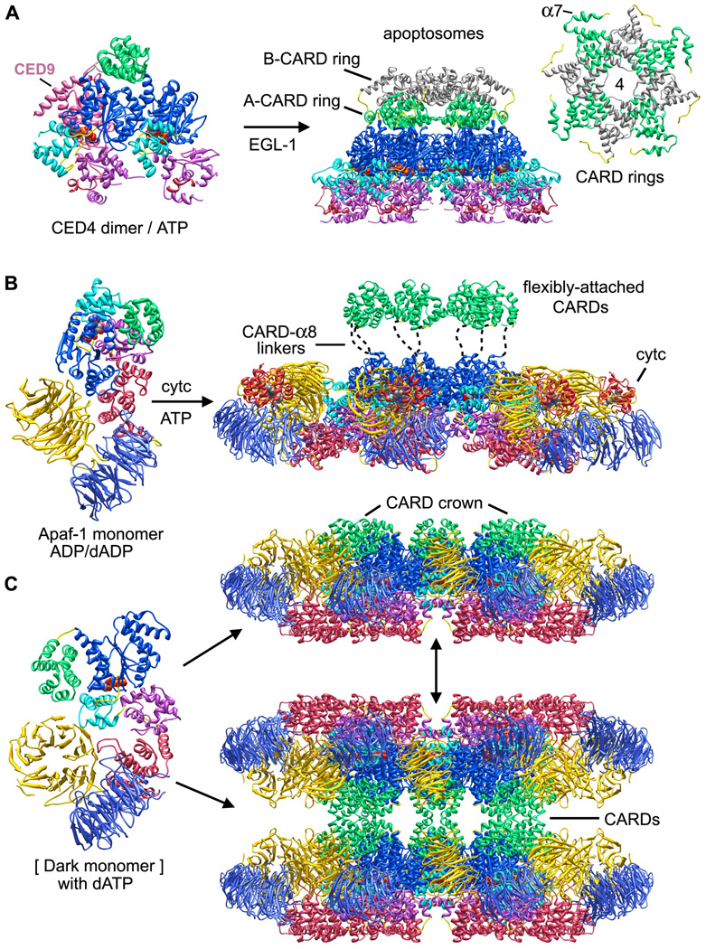
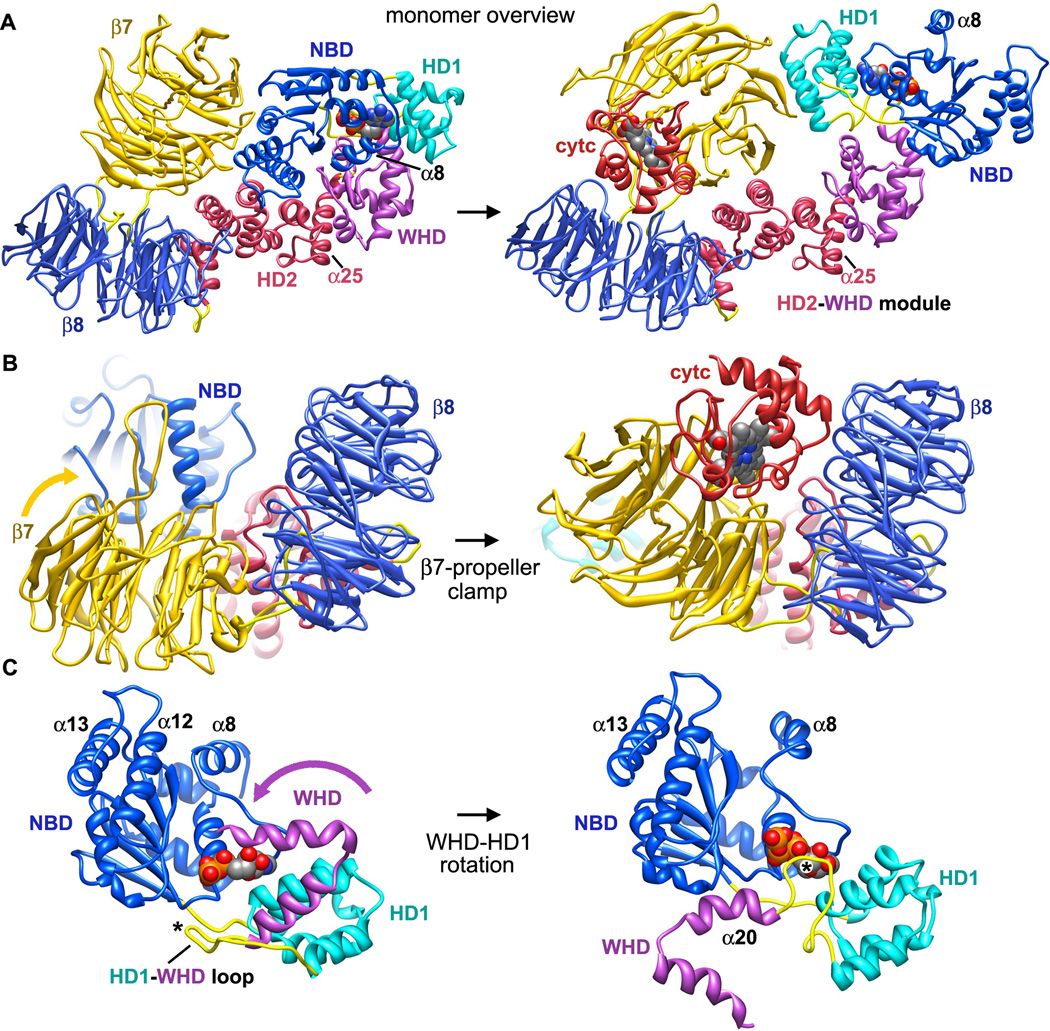
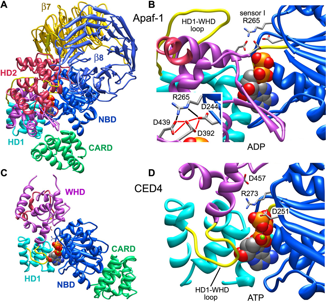
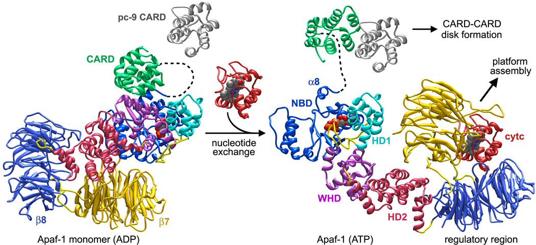
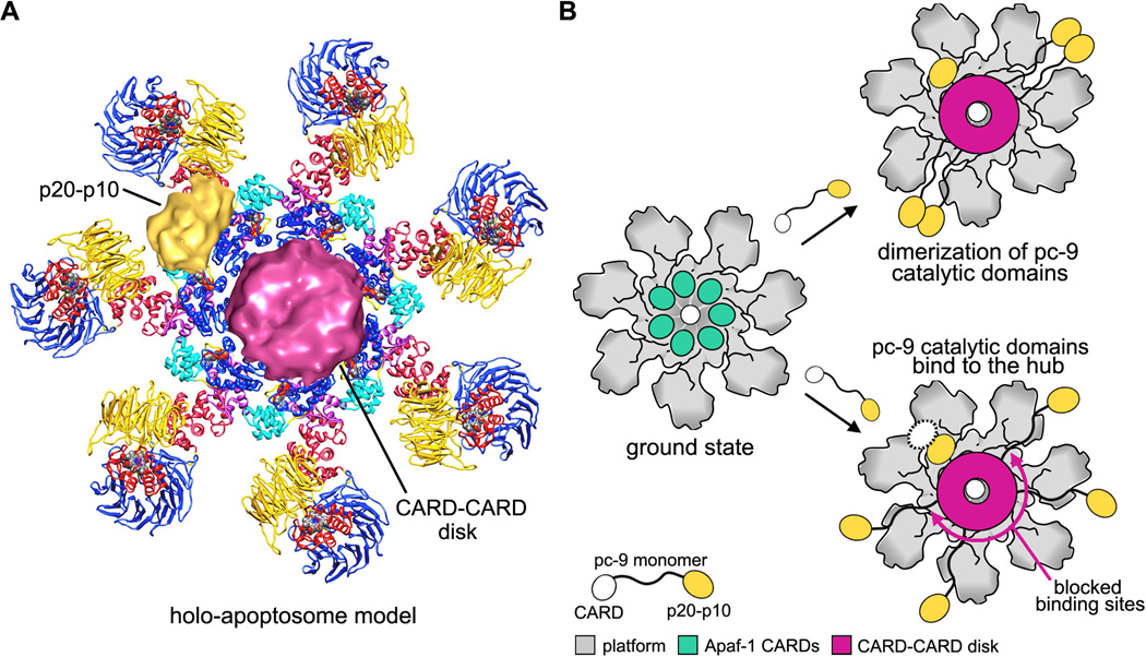
Apaf-1-like molecules assemble into a ring-like platform known as the apoptosome. This cell death platform then activates procaspases in the intrinsic cell death pathway. In this review, crystal structures of Apaf-1 monomers and CED-4 dimers have been combined with apoptosome structures to provide insights into the assembly of cell death platforms in humans, nematodes, and flies. In humans, the caspase recognition domains (CARDs) of procaspase-9 and Apaf-1 interact with each other to form a CARD-CARD disk, which interacts with the platform to create an asymmetric proteolysis machine. The disk tethers multiple pc-9 catalytic domains to the platform to raise their local concentration, and this leads to zymogen activation. These findings have now set the stage for further studies of this critical activation process on the apoptosome.


Figures

Figure 1
Figure 1. The intrinsic cell death pathway in worms, humans and flies. (left) In C. elegans, an apoptosome with quasi 8-fold symmetry assembles from lateral CED-4 dimers after they are released from an inhibited CED-9 complex by EGL-1. (middle) In humans, cytochrome c is released from mitochondria to trigger nucleotide exchange and assembly of a heptameric Apaf-1 apoptosome. (right) In Drosophila, Dark may bind dATP and assemble into active single rings or inactive double rings (not shown)
Figure 2
Figure 2. Domain architecture and structures of Apaf-1 like proteins in the NOD super-family
A. Linear domain representations are shown for CED-4, Apaf-1 and Dark. This domain color scheme is used throughout. B. A crystal structure is shown of the CED-9 inhibited CED-4 lateral dimer (2A5Y). The CARD in monomer B is disordered (Yan et al., 2005) and the boundary between monomers is indicated by a dashed line. C. A composite model is shown for an inactive Apaf-1 monomer based on two crystal structures (Reidl et al., 2005; Reubold et al., 2011).
Figure 3
Figure 3. Top views of worm, human and fly apoptosomes
A. (top left) The central hub of the C. elegans apoptosome has quasi 8-fold symmetry. (lower right) CARDs from the A and B monomers of CED-4 form two tetrameric rings stacked along the 8-fold axis of the hub to create an apoptosome with overall 4-fold symmetry (Qi et al., 2010; 3LQQ). B. The human apoptosome contains seven Apaf-1 molecules whose CARDs are disordered in the ground state (Yuan et al., 2010; submitted). C. A Drosophila single ring apoptosome is shown with eight CARDs that bind to the lateral surface of their respective NBDs to form a crown (Yuan et al., 2011a; 1VT4, 3IZ8). D. The CARD crown has been removed to show the similarity of the octagonal fly apoptosome and the heptameric Apaf-1 apoptosome in top views (compare with panel B). Note that mammalian cytochrome c does not bind to Dark in these complexes.
Figure 4
Figure 4. Assembly models for ground state apoptosomes in worms, humans and flies
A. CED-4 molecules in lateral dimers contain bound ATP and may represent an assembly intermediate blocked by CED-9 (Yan et al., 2005; Qi et al., 2010). EGL-1 interacts with CED-9 to promote further oligomerization of CED-4 to form the apoptosome. B. Inactive Apaf-1 monomers (Reidl et al. 2005; Reubold et al., 2011) bind cytochrome c and undergo nucleotide exchange to promote assembly of a heptameric apoptosome. CARDs are flexibly tethered to NBDs in the central hub by CARD-α8 linkers (Yuan et al., 2010). C. An extended Dark monomer was extracted from the apoptosome model and is shown on the left. In vitro assembly requires dATP to form single and double rings, as shown on the right.
Figure 5
Figure 5. Conformational transitions in Apaf-1 during apoptosome assembly
A. (left) A homology model of an inactive human Apaf-1 monomer with bound ADP is shown without the N-terminal CARD. (right) Cytochrome c binding to β-propellers induces a conformational change that may promote nucleotide exchange and assembly. Apaf-1 monomers have been aligned on their WHD-HD2 modules. The direction of view is approximately along the 7-fold axis of the apoptosome for the molecule on the right. B. The β7-propeller acts like a clamp to pin cytochrome c between the propellers. C. A close-up is shown of the rotation of WHD relative to the NBD and HD1 pair that occurs upon nucleotide exchange. The associated HD2 arm and β-propellers are not shown. The HD1-WHD loop (marked with an asterisk) with an S/TxYxY motif flips to interact with the ribose region of the bound nucleotide during, while the WHD-HD2 arm rotates. Helices α8, α12 and α13 also move during apoptosome assembly.
Figure 6
Figure 6. The role of sensor I in Apaf-1 monomer and CED4 apoptosomes
A. An icon view is shown of the Apaf-1 monomer. B. Stabilizing interactions of sensor I (R265) with an acidic triangle of carboxylate groups in the inactive Apaf-1 monomer are shown (see inset). Note the distal position of the HD1-WHD loop. C. An icon view is shown of a CED4 monomer extracted from the assembled apoptosome. D. Sensor I (R273) interactions are shown with the γ-phosphate of ATP in the CED-4 apoptosome. The HD1-WHD loop is in close proximity to the ribose of ATP.
Figure 7
Figure 7
Possible conformational changes that occur during Apaf-1 assembly are shown with inactive and active conformations aligned on the NBD. Cytochrome c binding may trigger inter-domain rearrangements and motions that lead to nucleotide exchange. An extensive rearrangement of the NBD-HD1 module occurs relative to the rest of Apaf-1. This creates an extended and assembly competent conformation. Procaspase-9 may interact with the CARD in the Apaf-1 monomer during nucleotide exchange leading directly to the formation of a CARD-CARD disk as the platform assembles.
Figure 8
Figure 8. Models of procaspase-9 activation on the human apoptosome
A. A composite model is shown for the holo-apoptosome, based on cryo-EM structures for the platform and active complex (Yuan et al. submitted). B. The platform is shown in grey and domains of pc-9 are color coded in a cartoon model of pc-9 activation. (top right) A proximity-induced dimerization model is shown with flexibly tethered pc-9 molecules. (bottom right) The proximity-induced association/allosteric model is shown. One p20-p10 catalytic domain (in gold) is bound to the hub and a possible second catalytic domain is shown as a dashed white oval, to indicate that it may be disordered.

References

    1. Acehan D, Jiang X, Morgan DG, Heuser JE, Wang X, Akey CW. Three-dimensional structure of the apoptosome: implications for assembly, procaspase-9 binding and activation. Mol. Cell. 2002;9:423–432. - PubMed
    1. Allan LA, Morrice N, Brady S, Magee G, Pathak S, Clarke PR. Inhibition of caspase-9 through phosphorylation at Thr 125 by ERK MAPK. Nat. Cell. Biol. 2003;5(7):647–654. - PubMed
    1. Allan LA, Clarke PR. Phosphorylation of caspase-9 by CDK1/cyclin B1 protects mitotic cells against apoptosis. Mol Cell. 2007;26:301–310. - PubMed
    1. Arama E, Bader M, Srivastava M, Bergmann A, Steller H. The two Drosophila cytochrome C proteins can function in both respiration and caspase activation. EMBO J. 2006;25:232–243. - PMC - PubMed
    1. Bao Q, Lu W, Rabinowitz JD, Shi Y. Calcium blocks formation of apoptosome by preventing nucleotide exchange in Apaf-1. Mol. Cell. 2007;25:181–192. - PubMed

Publication types

MeSH terms

LinkOut - more resources