

Hypomorphic mutations in PGAP2, encoding a GPI-anchor-remodeling protein, cause autosomal-recessive intellectual disability
- PMID: 23561846
- PMCID: PMC3617372
- DOI: 10.1016/j.ajhg.2013.03.008
Hypomorphic mutations in PGAP2, encoding a GPI-anchor-remodeling protein, cause autosomal-recessive intellectual disability
Abstract
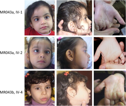
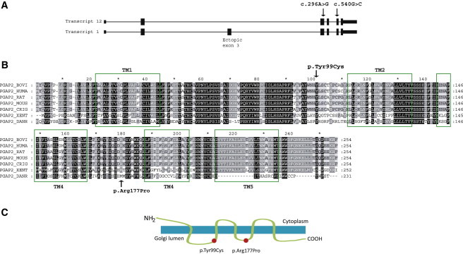
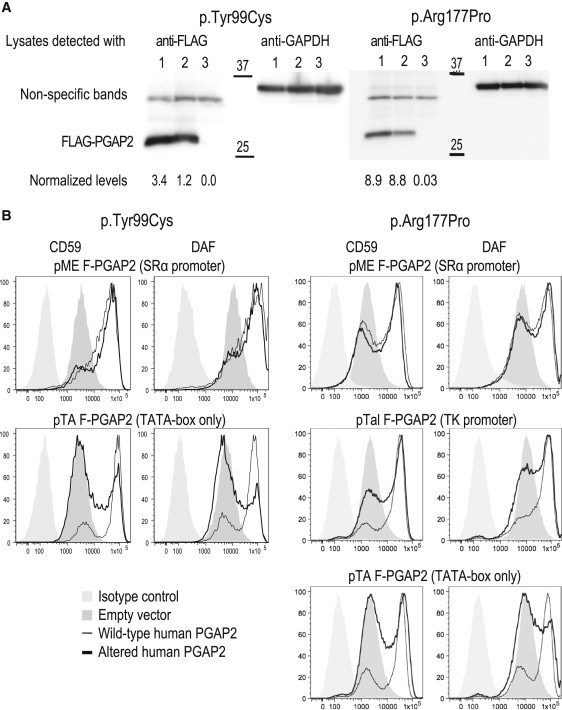
PGAP2 encodes a protein involved in remodeling the glycosylphosphatidylinositol (GPI) anchor in the Golgi apparatus. After synthesis in the endoplasmic reticulum (ER), GPI anchors are transferred to the proteins and are remodeled while transported through the Golgi to the cell membrane. Germline mutations in six genes (PIGA, PIGL, PIGM, PIGV, PIGN, and PIGO) in the ER-located part of the GPI-anchor-biosynthesis pathway have been reported, and all are associated with phenotypes extending from malformation and lethality to severe intellectual disability, epilepsy, minor dysmorphisms, and elevated alkaline phosphatase (ALP). We performed autozygosity mapping and ultra-deep sequencing followed by stringent filtering and identified two homozygous PGAP2 alterations, p.Tyr99Cys and p.Arg177Pro, in seven offspring with nonspecific autosomal-recessive intellectual disability from two consanguineous families. Rescue experiments with the altered proteins in PGAP2-deficient Chinese hamster ovary cell lines showed less expression of cell-surface GPI-anchored proteins DAF and CD59 than of the wild-type protein, substantiating the pathogenicity of the identified alterations. Furthermore, we observed a full rescue when we used strong promoters before the mutant cDNAs, suggesting a hypomorphic effect of the mutations. We report on alterations in the Golgi-located part of the GPI-anchor-biosynthesis pathway and extend the phenotypic spectrum of the GPI-anchor deficiencies to isolated intellectual disability with elevated ALP. GPI-anchor deficiencies can be interpreted within the concept of a disease family, and we propose that the severity of the phenotype is dependent on the location of the altered protein in the biosynthesis chain.
Copyright © 2013 The American Society of Human Genetics. Published by Elsevier Inc. All rights reserved.
Figures
References
-
- Kinoshita T., Fujita M., Maeda Y. Biosynthesis, remodelling and functions of mammalian GPI-anchored proteins: recent progress. J. Biochem. 2008;144:287–294. - PubMed
-
- Fujita M., Kinoshita T. GPI-anchor remodeling: potential functions of GPI-anchors in intracellular trafficking and membrane dynamics. Biochim. Biophys. Acta. 2012;1821:1050–1058. - PubMed
-
- Maeda Y., Kinoshita T. Structural remodeling, trafficking and functions of glycosylphosphatidylinositol-anchored proteins. Prog. Lipid Res. 2011;50:411–424. - PubMed
Publication types
MeSH terms
Substances
LinkOut - more resources
Full Text Sources
Other Literature Sources
Medical
Molecular Biology Databases
Miscellaneous
