. 2013 Jul:232:76-86.
doi: 10.1016/j.jmr.2013.02.014. Epub 2013 Mar 14.

Diffusion weighted MRI by spatiotemporal encoding: analytical description and in vivo validations

Affiliations

Diffusion weighted MRI by spatiotemporal encoding: analytical description and in vivo validations

Eddy Solomon et al. J Magn Reson. 2013 Jul.

Abstract

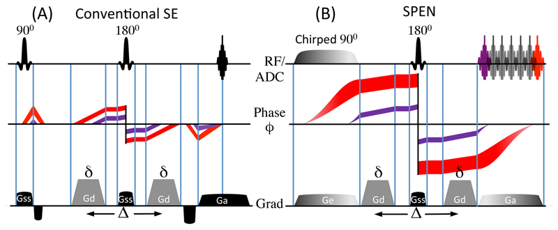
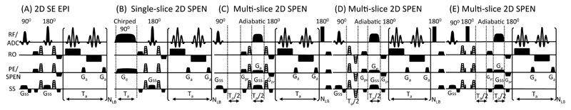
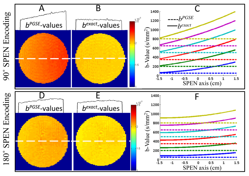
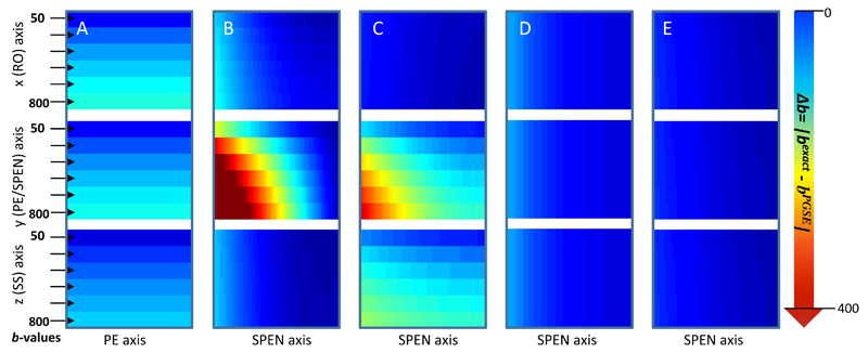
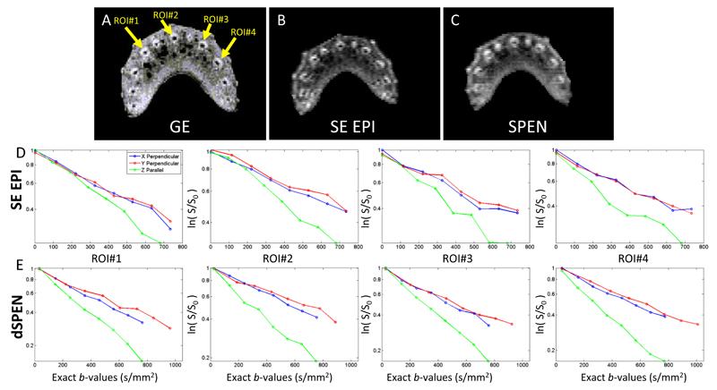
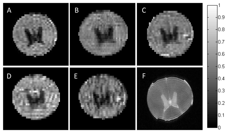
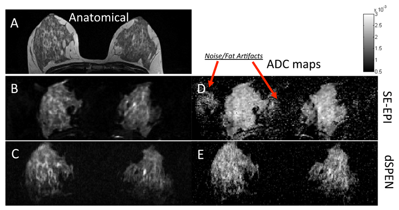
Diffusion-weighted (DW) MRI is a powerful modality for studying microstructure in normal and pathological tissues. The accuracy derived from DW MRI depends on the acquisition of quality images, and on a precise assessment of the b-values involved. Conventional DW MRI tends to be of limited use in regions suffering from large magnetic field or chemical shift heterogeneities, which severely distort the MR images. In this study we propose novel sequences based on SPatio-temporal ENcoding (SPEN), which overcome such shortcomings owing to SPEN's inherent robustness to offsets. SPEN, however, relies on the simultaneous application of gradients and radiofrequency-swept pulses, which may impart different diffusion weightings along the spatial axes. These will be further complicated in DW measurements by the diffusion-sensitizing gradients, and will in general lead to complex, spatially-dependent b-values. This study presents a formalism for analyzing these diffusion-weighted SPEN (dSPEN) data, which takes into account the concomitant effects of adiabatic pulses, of the imaging as well as diffusion gradients, and of the cross-terms between them. These analytical b-values derivations are subject to experimental validations in phantom systems and ex vivo spinal cords. Excellent agreement is found between the theoretical predictions and these dSPEN experiments. The ensuing methodology is then demonstrated by in vivo mapping of diffusion in human breast - organs where conventional k-space DW acquisition methods are challenged by both field and chemical shift heterogeneities. These studies demonstrate the increased robustness of dSPEN vis-à-vis comparable DW echo planar imaging, and demonstrate the value of this new methodology for medium- or high-field diffusion measurements in heterogeneous systems.


Figures

Figure 1
Figure 1
1D Spin-Echo (A) and 1D SPEN (B) imaging experiments, depicting the phase dispersion undergone by two spin packets in each sequence. Whereas in 1D SE all spins experience the same local dephasing throughout the sample, in SPEN, the quadratic dependence (Eq. 2) induce different local ϕ-dispersion. The cartoons illustrates this for two spin-packets colored in red and purple; as b-values depend on the degree of local dephasing, a spatial weighting throughout the FOV is incurred. The RF/ADC line displays the RF and signal acquisition timings; the gradients are: Ge = encoding gradient; Gd = diffusion gradient; Gss = slice-selective gradient; Ga = acquisition gradient; diffusion timing parameters are δ and ∆.
Figure 2
Figure 2
2D single-scan diffusion pulse sequences assayed, with their gradients and timing definitions (delays not drawn to scale). In the 2D Spin-Echo EPI (A), single-slice 2D SPEN (B) and multi-slice 2D SPEN (C) sequences, the diffusion-sensitizing gradients are located on both sides of a central refocusing pulse. In (D) the diffusion blocks is placed in a Ta/2 delay required by SPEN’s full refocusing procedure; in (E) Gd operates as a separate diffusion block in combination with a 180° hard pulse, leading to a compensation of cross-terms and reduced spatial b-dephasing. All SPEN diffusion sequences are fully refocused. The RF/ADC line displays the RF and signal acquisition; other definitions: Ge = encoding gradient; Gd = diffusion gradient (shown in gray for all sequences); Gss = slice-selective gradient; Gpr = purge gradient; Ga = acquisition gradient; Ta = acquisition time; NLB = number of loops encoding the low-bandwidth (PE/SPEN) dimensions.
Figure 3
Figure 3
Diffusion results for pure H2O using 90° chirp (top panels) and 180° adiabatic sweep (lower panels) 2D SPEN sequences, upon ignoring (A, D) or accounting (B, E) for the effects of SPEN on the b-values. The A,B and D,E panels present the ADC maps together with 1D profiles illustrating cross-sections taken along the SPEN dimensions at the dashed white lines. The graphs in C, F compare the b-values expected solely on the basis of PGSE calculation (dashed lines) against the exact calculations as derived in Section 2 (solid lines). The diffusion parameters are δ = 3 ms, ∆ = 14 ms. The scanning parameters: TR=5sec, cubic FOV = 30x30 mm2, resolution = 0.4×0.4 mm2 on a 2 mm slice, number of averages = 4. Gradient and timing values for the 90° SPEN experiments: T90 = 21ms, Ge = 1.2 G/cm, Ta = 21ms, Ga = 4.5 G/cm, total scan duration = 60 ms. Idem for the 180° SPEN parameters: T90 = 2 ms, T180 = 10.5 ms, Ge = 0.8 G/cm, Ta = 21 ms, Ga = 3 G/cm and a total scan duration = 51ms.
Figure 4
Figure 4
Residual∆b-values reflecting |bexact(including all imaging gradients) – bPGSE (eq.1b)|, for the five diffusion experiments described in Fig. 2. The columns correspond one-to-one to these sequences lettering, and the rows correspond to diffusion gradients applied along the indicated axes. Notice the minimal disturbance evidenced by the sequences in Fig. 2D, 2E. Parameters assumed for these diffusion calculations are δ= 3ms, ∆ = 14ms (except for Fig. 4D where ∆ = 11.4ms). The gradient and timing values taken into account for the b-value calculation are according to the experimental scanning parameters presented in Fig. 3 and Fig. B1 of Appendix B
Figure 5
Figure 5
Magnitude gradient-echo (GE, A), single-scan SE EPI (B) and single-scan SPEN (C) images, displaying the different vascular bundles in fresh celery. Diffusion curves of SE EPI and dSPEN measurements at the center of the vascular bundles measured parallel (along the Z) and perpendicular (along X and Y) to it, are shown in (D) and (E) respectively. Each plot shows the average log of signal intensity vs bexact-values for four different vascular ROI’s (see arrows in A). The diffusion parameters are δ = 10ms and ∆ = 20ms. SE EPI and SPEN scanning parameters: FOV = 28 x 22 mm2, nominal resolution = 0.4×0.31 mm2 on a 3 mm slice. For the multi-scan GE: FOV = 20x20 mm2, resolution = 0.15×0.15 mm2 on a 3 mm slice total scan duration = 4 sec. Gradient and timing values for SE EPI: T90=T180=3ms, Ta=21ms, total scan duration = 67ms. For SPEN: T90 = 3 ms, Ge = 10 G/cm, Ta = 21 ms, Ga = 5.3 G/cm, total scan duration = 57 ms.
Figure 6
Figure 6
(A-E) FA maps of ex-vivo swine spinal cord scanned by the pulse sequences described in Fig. 2 –panels A-E respectively. (F) Anatomical multi-scan fast spin-echo (FSE) reference image. The scale bar for the FA maps runs from 0 for isotropic to 1 for fully anisotropic diffusion. Maps were Generated considering the readout, phase-encode and slice-selection directions as λ1, λ2, λ3 respectively. Decay curves employed bexact-values as described in Section 2. Diffusion parameters are δ= 3ms, ∆ = 14ms (except for Fig. 6D where ∆ = 11.4ms) weighted by bPGSE-values: 50 250 450 650 850 1050. In all cases, corrected b-values were used to derive the maps. Scanning parameters of (A-E) are: TR=5sec, cubic FOV = 2 x20 mm2, nominal resolution = 0.28×0.28 mm2 on a 2 mm slice, number of averages = 4. (F) FOV = 20x20 mm2, resolution = 0.1×0.1 mm2 on a 1 mm slice, total scan duration = 2.5 sec. Other parameters were as follows: (A) T90=T180=2ms, Ta=21ms, total duration = 51.5ms. (B) T90=Ta=21 ms, Ge=1.2 G/cm, Ga=4.5 G/cm, total scan duration = 60ms. (C) T90=2ms, T180=Ta/2=10.5ms, Ge=0.8 G/cm, Ga=3 G/cm, total scan duration = 51ms. (D) T90=2ms, T180=Ta/2=15.4ms, Ge=0.4 G/cm, Ga=4.4 G/cm, total scan duration = 65ms. (E) T90=2ms, T180=Ta/2=10.5ms, Ge=0.8 G/cm, Ga=3 G/cm, total scan duration = 62ms.
Figure 7
Figure 7
Comparison between anatomical images (left-hand panels) and ADC maps (right) collected on a human breast-scan volunteer. (A) Multi-scan T2 turbo spin echo image. (B) Axial slice, twice-refocused EPI. (C) Single-slice SPEN (sequence in Fig. 2B). The corresponding ADC maps (D, E) were originally weighted by bPGSE-values 0 250 500 750 1000, arising from δ= 26.3 ms, ∆ = 43.4 ms. In the SPEN case, corrected b-values were used to derive the maps. Scanning parameters for the multi-scan image: cubic FOV= 360 mm on each side; resolution = 0.8×0.6 mm2 on a 2.5 mm slice; total scan duration = 1080ms (without fat suppression). For EPI: square FOV = 360x360 mm2, resolution = 1.9×1.9 mm2, 2.5 mm slice, total scan duration (with GRAPPA parallel imaging, not used in the SPEN case) = 150 ms. SPEN parameters: FOV = 300×120 mm2, resolution = 1.6×1.6 mm2 on a 2.5 mm slice, T90 = 47.8 ms, Ge = 0.06 G/cm, Ta = 66.1 ms and Ga = 0.043 G/cm, total scan duration = 200ms . For both EPI and SPEN, fat suppression was used.

References

    1. Basser PJ, Jones DK. Diffusion-tensor MRI: theory, experimental design and data analysis - a technical review. NMR Biomed. 2002;15:456–467. - PubMed
    1. Callaghan PT. Principles of nuclear magnetic resonance microscopy. Corrected ed. Clarendon Press; Oxford: 1993.
    1. Le Bihan D, Mangin JF, Poupon C, Clark CA, Pappata S, Molko N, Chabriat H. Diffusion tensor imaging: Concepts and applications. J Magn Reson. 2001;13:534–546. - PubMed
    1. Norris DG, Driesel W. Online motion correction for diffusion-weighted imaging using navigator echoes: Application to RARE imaging without sensitivity loss. Magn Reson Med. 2001;45:729–733. - PubMed
    1. Nunes RG, Jezzard P, Behrens TE, Clare S. Self-navigated multishot echo-planar pulse sequence for high-resolution diffusion-weighted imaging. Magn Reson Med. 2005;53:1474–1478. - PubMed

Publication types

LinkOut - more resources