. 2013 Jun;39(6):1102-19.
doi: 10.1016/j.ultrasmedbio.2013.01.013. Epub 2013 Apr 3.

In vitro gene delivery with ultrasound-triggered polymer microbubbles

Affiliations

In vitro gene delivery with ultrasound-triggered polymer microbubbles

Michael Cochran et al. Ultrasound Med Biol. 2013 Jun.

Abstract

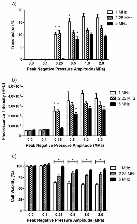
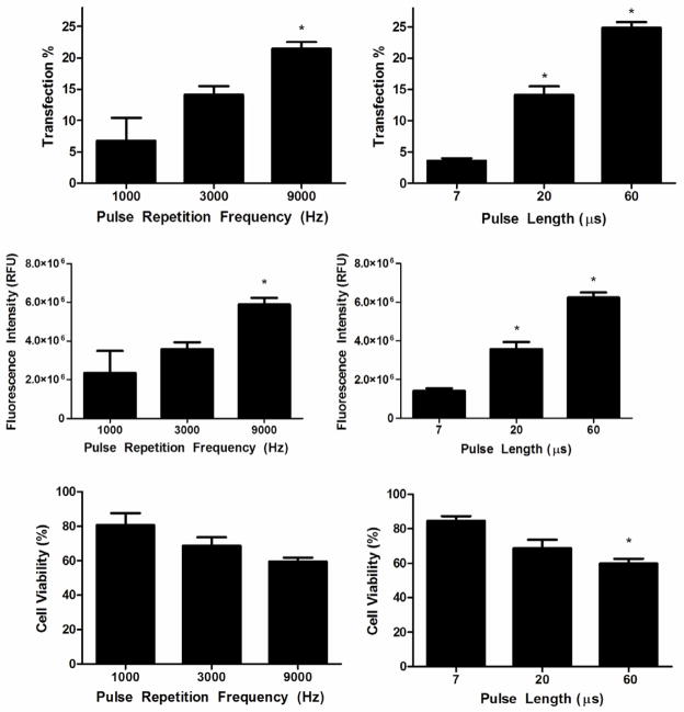
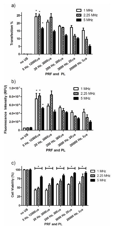
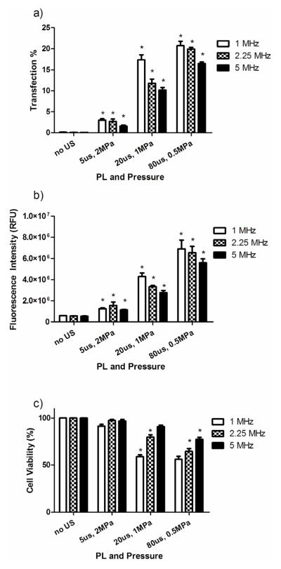
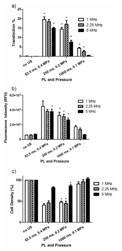
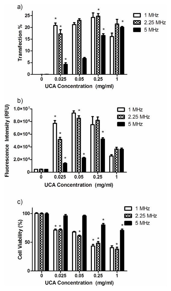
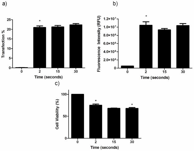
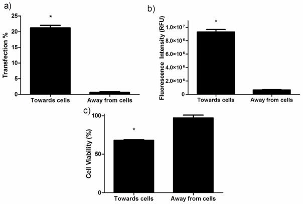
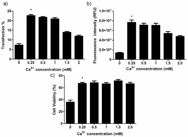
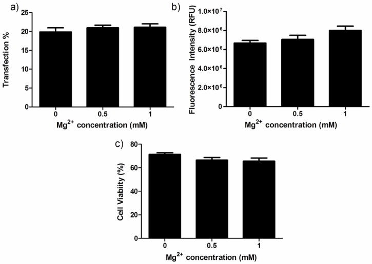
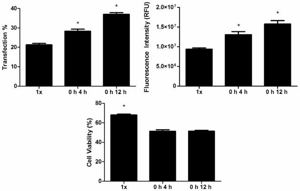
In the work described here, gene delivery using polymer microbubbles triggered by ultrasound in vitro was investigated. The effects of pressure amplitude (0-2 MPa), center frequency (1-5 MHz), pulse length (3-12,000 μs), pulse repetition frequency (5-20,000 Hz) and exposure time (0-30 s) on transfection efficiency and cell viability were examined. The effects of radiation force, calcium ion concentration and timing of treatments were also examined. Cells were successfully transfected with pressure amplitudes as low as 250 kPa. Transfection was most efficient at lower frequencies and longer pulse lengths, with a transfection efficiency of 24.2 ± 2.0% achieved using a center frequency of 1 MHz, pressure amplitude of 1 MPa, pulse length of 12,000 μs and pulse repetition frequency of 5 Hz. Gene delivery was also affected by the extracellular calcium ion concentration and the timing of treatments.


Figures

Figure 1
Figure 1
The effect of pressure amplitude and center frequency on (a) transfection efficiency, (b) total fluorescence intensity and (c) cell viability. MCF 7 cells were insonated for 15 seconds with 0.25 mg/ml PLA UCA and 10 μg/ml plasmid DNA with a PL of 20 μs and PRF of 3000 Hz (*p<0.05, n=5).
Figure 2
Figure 2
The effect of pulse repetition frequency on (a) transfection efficiency, (b) total fluorescence intensity and (c) viability of cells insonated with a constant pulse length of 20 μs and the effect of pulse length on (d) transfection efficiency, (e) total fluorescence intensity and (f) viability of cells insonated with a constant pulse repetition frequency of 3000 Hz. All cells were insonated for 15 seconds at 1 MHz with a pressure amplitude of 500 kPa and a PLA microbubble concentration of 0.25 mg/ml (*p<0.05, n=4).
Figure 3
Figure 3
The effect of pulse repetition frequency and pulse length on (a) transfection efficiency, (b) total fluorescence intensity and (c) cell viability. Cells were insonated for 15 seconds with 0.25 mg/ml PLA UCA and 10 μg/ml plasmid DNA with a pressure amplitude of 1 MPa and a constant duty cycle of 0.06 (*p<0.05, n=5).
Figure 4
Figure 4
Fluorescent images of MCF 7 breast cancer cells expressing GFP 24 hours after exposure to ultrasound with a constant acoustic pressure amplitude of 1 MPa while the PRF and PL were simultaneously adjusted to maintain a constant DC of 0.06 and an ISPTA of 2.0 W/cm2 (size bar = 100 μm).
Figure 5
Figure 5
Effect of PL and pressure amplitude on (a) transfection efficiency, (b) total fluorescence intensity and (c) viability of cells insonated with a constant PRF of 3000 Hz and a pressure and PL that were simultaneously changed to maintain a constant ISPTA of 2.0 W/cm2 (*p<0.05, n=5).
Figure 6
Figure 6
Effect of PL and pressure amplitude on (a) transfection efficiency, (b) total fluorescence intensity and (c) viability of cells insonated for 15 seconds with a constant PRF of 1 Hz and a pressure and PL that were simultaneously changed to maintain a constant ISPTA of 0.33 W/cm2 (*p<0.05, n=5).
Figure 7
Figure 7
The effect of PLA microbubble concentration on (a) transfection efficiency, (b) total fluorescence intensity and (c) viability of cells insonated for 15 seconds with a constant pressure amplitude of 1 MPa, PL of 12 ms and PRF of 5 Hz (*p<0.05, n=5).
Figure 8
Figure 8
The effect of insonation time on the (a) transfection efficiency, (b) total fluorescence intensity and (c) cell viability of cells insonated with 0.05 mg/ml UCA and a center frequency of 1 MHz, pressure amplitude of 1 MPa, PL of 12 ms and PRF of 5 Hz (*p<0.05, n=6).
Figure 9
Figure 9
The effect of radiation forces on (a) transfection efficiency, (b) total fluorescence intensity and (c) cell viability of cells insonated for 15 seconds with a center frequency of 1 MHz, a pressure amplitude of 1 MPa, PL of 12 ms, PRF of 5 Hz and UCA concentration of 0.05 mg/ml. The direction of ultrasound exposure was changed from pushing UCA towards cells to away from cells (*p<0.05, n=6).
Figure 10
Figure 10
The effect of calcium ion concentration on (a) transfection efficiency, (b) total fluorescence intensity and (c) cell viability of cells insonated for 15 seconds with a center frequency of 1 MHz, a pressure amplitude of 1 MPa, PL of 12 ms, PRF of 5 Hz and UCA concentration of 0.05 mg/ml. Microbubbles and plasmid were suspended in PBS containing 0.49 mM magnesium chloride and 0–2 mM calcium chloride (*p<0.05, n=6).
Figure 11
Figure 11
The effect of magnesium ion concentration on (a) transfection efficiency, (b) total fluorescence intensity and (c) cell viability of cells insonated for 15 seconds with a center frequency of 1 MHz, a pressure amplitude of 1 MPa, PL of 12 ms, PRF of 5 Hz and UCA concentration of 0.05 mg/ml. Microbubbles and plasmid were suspended in PBS containing 1.0 mM calcium chloride and 0–1 mM magnesium chloride (n=6).
Figure 12
Figure 12
The effect of multiple exposures on (a) transfection efficiency, (b) total fluorescence intensity and (c) cell viability of cells insonated for 15 seconds with 0.05 mg/ml UCA at a center frequency of 1 MHz, a pressure amplitude of 1 MPa, PL of 12 ms and PRF of 5 Hz. Cells were treated either once (1x) or twice, with the second exposure 4 hours after the first (0 h 4 h) or 12 hours after the first (0 h 12 h) (*p<0.05, n=5).

Similar articles

Cited by

References

    1. Apfel RE, Holland CK. Gauging the likelihood of cavitation from short-pulse, low-duty cycle diagnostic ultrasound. Ultrasound Med Biol. 1991;17:179–85. - PubMed
    1. Azmin M, Harfield C, Ahmad Z, Edirisinghe M, Stride E. How Do Microbubbles and Ultrasound Interact? Basic Physical, Dynamic and Engineering Principles. Curr Pharm Des. 2012;18:2118–34. - PubMed
    1. Balazs DA, Godbey W. Liposomes for Use in Gene Delivery. Journal of Drug Delivery. 2011 - PMC - PubMed
    1. Bao S, Thrall BD, Miller DL. Transfection of a reporter plasmid into cultured cells by sonoporation in vitro. Ultrasound Med Biol. 1997;23:953–9. - PubMed
    1. Bohmer MR, Chlon CHT, Raju BI, Chin CT, Shevchenko T, Klibanov AL. Focused ultrasound and microbubbles for enhanced extravasation. J Control Release. 2010;148:18–24. - PMC - PubMed

Publication types