. 2013 Apr 2;17(4):549-61.
doi: 10.1016/j.cmet.2013.03.009.

Impaired cholesterol efflux in senescent macrophages promotes age-related macular degeneration

Affiliations

Impaired cholesterol efflux in senescent macrophages promotes age-related macular degeneration

Abdoulaye Sene et al. Cell Metab. .

Abstract

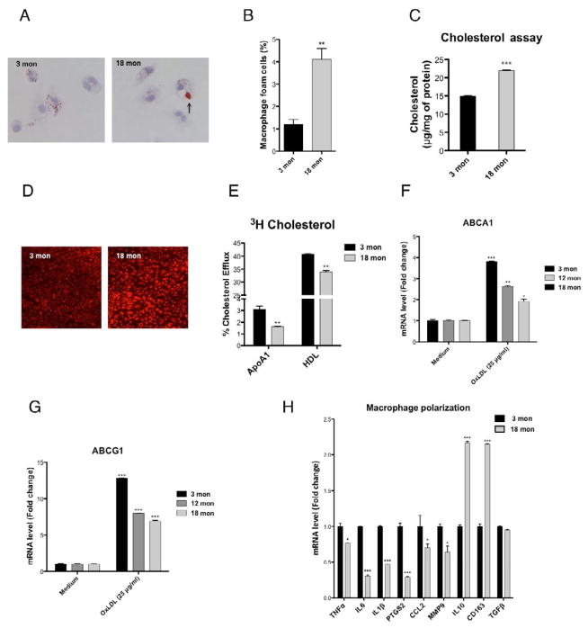
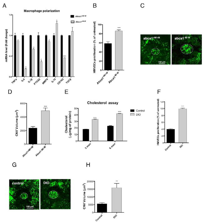
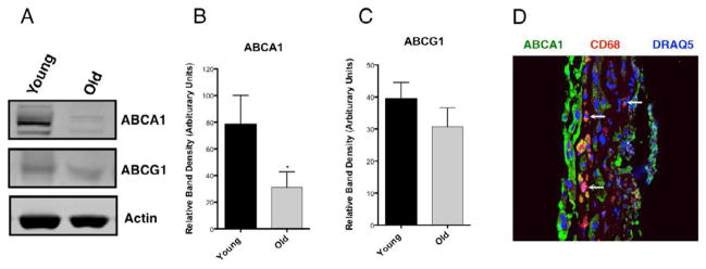
Pathologic angiogenesis mediated by abnormally polarized macrophages plays a central role in common age-associated diseases such as atherosclerosis, cancer, and macular degeneration. Here we demonstrate that abnormal polarization in older macrophages is caused by programmatic changes that lead to reduced expression of ATP binding cassette transporter ABCA1. Downregulation of ABCA1 by microRNA-33 impairs the ability of macrophages to effectively efflux intracellular cholesterol, which in turn leads to higher levels of free cholesterol within senescent macrophages. Elevated intracellular lipid polarizes older macrophages to an abnormal, alternatively activated phenotype that promotes pathologic vascular proliferation. Mice deficient for Abca1, but not Abcg1, demonstrate an accelerated aging phenotype, whereas restoration of cholesterol efflux using LXR agonists or miR-33 inhibitors reverses it. Monocytes from older humans with age-related macular degeneration showed similar changes. These findings provide an avenue for therapeutic modulation of macrophage function in common age-related diseases.


Figures

Figure 1
Figure 1. ABCA1 and ABCG1 expression is altered in senescent macrophages
Quantitative mRNA analysis of ABCA1 and ABCG1 in splenic (A), peritoneal (B) macrophages and PBMCs (C) isolated from 3, 12 or 18 months old mice. (D) CNV was induced in 3 or 18 months old mice and eye macrophages harvested by laser microdissection were analyzed for ABCA1 and ABCG1 expression by qPCR. (E) Whole cell lysate of peritoneal macrophages of 3 or 18 months old mice were subjected to immunoblot analysis with antibodies against ABCA1, ABCG1 or β-Actin. (F) Relative flow cytometry profiles of ABCA1 and ABCG1 in peritoneal macrophages isolated from 3 or 18 months old mice. Iso = negative isotype control. Values are expressed as mean + SE (A–D). Statistically significant difference *P<0.05, **P<0.01, ***P<0.001.
Figure 2
Figure 2. Age associated impairment in macrophage cholesterol efflux capacities
(A) Thioglycollate elicited young or old peritoneal macrophages were stained with Oil Red O and hematoxylin and (B) quantitative analysis of foamy macrophages was performed. (C) Total cholesterol content of young or old macrophage was measured and normalized to protein content. (D) Peritoneal macrophages were incubated with DiI-oxLDL and intracellular accumulation of oxLDL was assessed by confocal microscopy. (E) Cholesterol efflux to ApoA1 and HDL of peritoneal macrophages pre-loaded with [3H]cholesterol-oxLDL. Peritoneal macrophages isolated from 3, 12 or 18 months old mice were treated with 25 μg/ml oxLDL and relative mRNA expression of ABCA1 (F) and ABCG1 (G) was determined. (H) Young and old macrophages polarization. Expression of M1 (TNFα, IL6, ILlβ, PTGS2, CCL2 and MMP9) and M2 (IL10, CD163 and TGFβ) markers was analyzed by qPCR. Values are expressed as mean + SD (B) and mean + SE (C, E–H). Statistically significant difference *P<0.05, **P<0.01, ***P<0.001.
Figure 3
Figure 3. Macrophage ability to regulate vascular proliferation is altered by Abca1 deletion or cholesterol-rich diet
(A) Quantitative mRNA analysis of M1 (TNFα, IL6, ILlβ, PTGS2, CCL2 and MMP9) and M2 (IL10, CD163 and TGFβ) markers in wt (Abca1+M/+M) and ABCA1-deficient (Abca1M/M) peritoneal macrophages. (B) HMVECs proliferation when co-cultured with Abca1+M/+M or Abca1M/M macrophages. (C) Abca1+M/+M or Abca1M/M mice were perfused with FITC-dextran (green) 7 days after laser injury and CNV was examined by confocal microscopy. Representative CNV photographs are shown, scale bar; 100 μm. (D) Quantification of CNV (white circle) demonstrated a significant increase of CNV volume in Abca1M/M mice compared to wt mice. (E) Quantification of relative cholesterol content demonstrated a higher accumulation in macrophages from DIO mice at both 3 and 6 months of age as compared to controls. (F) Measurement of proliferation of HMVECs after incubation with DIO or control macrophages. Representative illustration (G) and quantification of CNV (white circle) volume (H) demonstrated an inability of DIO macrophages to inhibit CNV as compared to controls. Values are expressed as mean + SE. Statistically significant difference **P<0.01, ***P<0.001.
Figure 4
Figure 4. LXR agonist treatment restored the functional capacities of senescent macrophages
Peritoneal macrophages isolated from young and old mice were treated with vehicle, 1nM to 10 μM of T0-901317 (T0) and quantitative analysis of ABCA1 (A) and ABCG1 (B) gene expression was performed by real time qPCR. (C) Oxysterols (27-hydroxycholesterol (27-HC) and 7-ketocholesterol (7-KC)) extracted from culture medium of young or old peritoneal macrophages were quantified by HPLC/MS and normalized to macrophage protein content. (D) Effect of T0-901317 treatment on the ability of macrophages to inhibit HMVECs proliferation. Quantitative mRNA levels of ABCA1 and ABCG1 in retina (E), choroid (F) and peritoneal macrophages (G) of old mice, treated for 5 consecutive days with vehicle, 25 mg/kg or 50 mg/kg of T0-901317. (H) Representative CNV images in vehicle and LXR agonist treated old mice and quantification (I) of CNV (white circle) volume show a dose dependent reduction of endothelial cell proliferation. Values are expressed as mean + SE. Statistically significant difference *P<0.05, **P<0.01, ***P<0.001, compared to vehicle treatment.
Figure 5
Figure 5. Age-related alteration of ABCA1 expression in human PBMCs
(A) Western blot analysis of proteins extracted from PBMCs of young (< 35 years old) or old (> 65 years old) donors. Immunoblots were probed with antibodies against ABCA1, ABCG1 or β-Actin. Relative quantification of ABCA1 (B) and ABCG1 (C) band densities. (D) Immunohistochemistry of a CNV sample isolated from a patient after sub-retinal surgery with antibodies against ABCA1 (green) or CD68 (macrophage marker, red). Nuclei were stained with Draq5 (blue). Values are expressed as mean + SE. Statistically significant difference *P<0.05.
Figure 6
Figure 6. MiR-33 modulates macrophage regulation of vascular proliferation
(A) Quantitative analysis of miR-33 in young and old peritoneal macrophage. (B) Representative photograph of macrophages untransfected (UT) or transfected with fluorescent LNA anti-miR. Macrophages isolated from 3 or 18 months old mice were transfected with negative control anti-miR or anti-miR-33. 24 hours after transfection, mRNA (C) and protein (D) expression of ABCA1 was determined. (E) Effects of miR-33 antagonism on macrophage inhibition of HMVECs proliferation. Old mice were daily pre-treated with 10 mg/kg in vivo LNA, negative control anti-miR or anti-miR-33 for 7 days prior induction of CNV. Mice were then treated for 7 additional days and quantitative mRNA (F) and protein (G) level of ABCA1 in the choroid was analyzed. Representative illustration (H) and CNV volume quantification (I) showed that in vivo antagonism of miR-33 induced an inhibition of vascular endothelial cell proliferation. Values are expressed as mean + SE. Statistically significant difference *P<0.05, **P<0.01.

Comment in

References

    1. Apte RS, Richter J, Herndon J, Ferguson TA. Macrophages inhibit neovascularization in a murine model of age-related macular degeneration. PLoS Med. 2006;3:e310. - PMC - PubMed
    1. Baldan A, Tarr P, Vales CS, Frank J, Shimotake TK, Hawgood S, Edwards PA. Deletion of the transmembrane transporter ABCG1 results in progressive pulmonary lipidosis. J Biol Chem. 2006;281:29401–29410. - PubMed
    1. Bressler SB, Maguire MG, Bressler NM, Fine SL. Relationship of drusen and abnormalities of the retinal pigment epithelium to the prognosis of neovascular macular degeneration. The Macular Photocoagulation Study Group. Arch Ophthalmol. 1990;108:1442–1447. - PubMed
    1. Chen W, Stambolian D, Edwards AO, Branham KE, Othman M, Jakobsdottir J, Tosakulwong N, Pericak-Vance MA, Campochiaro PA, Klein ML, Tan PL, Conley YP, Kanda A, Kopplin L, Li Y, Augustaitis KJ, Karoukis AJ, Scott WK, Agarwal A, Kovach JL, Schwartz SG, Postel EA, Brooks M, Baratz KH, Brown WL, Brucker AJ, Orlin A, Brown G, Ho A, Regillo C, Donoso L, Tian L, Kaderli B, Hadley D, Hagstrom SA, Peachey NS, Klein R, Klein BE, Gotoh N, Yamashiro K, Ferris F, III, Fagerness JA, Reynolds R, Farrer LA, Kim IK, Miller JW, Corton M, Carracedo A, Sanchez-Salorio M, Pugh EW, Doheny KF, Brion M, Deangelis MM, Weeks DE, Zack DJ, Chew EY, Heckenlively JR, Yoshimura N, Iyengar SK, Francis PJ, Katsanis N, Seddon JM, Haines JL, Gorin MB, Abecasis GR, Swaroop A. Genetic variants near TIMP3 and high-density lipoprotein-associated loci influence susceptibility to age-related macular degeneration. Proc Natl Acad Sci U S A. 2010;107:7401–7406. - PMC - PubMed
    1. Chakravarthy MV, Pan Z, Zhu Y, Tordjman K, Schneider JG, Coleman T, Turk J, Semenkovich CF. “New” hepatic fat activates PPARalpha to maintain glucose, lipid, and cholesterol homeostasis. Cell Metab. 2005;1:309–322. - PubMed

Publication types

MeSH terms