. 2013 Apr 2;17(4):607-17.
doi: 10.1016/j.cmet.2013.03.004.

Tanycytic VEGF-A boosts blood-hypothalamus barrier plasticity and access of metabolic signals to the arcuate nucleus in response to fasting

Affiliations

Tanycytic VEGF-A boosts blood-hypothalamus barrier plasticity and access of metabolic signals to the arcuate nucleus in response to fasting

Fanny Langlet et al. Cell Metab. .

Abstract

The delivery of blood-borne molecules conveying metabolic information to neural networks that regulate energy homeostasis is restricted by brain barriers. The fenestrated endothelium of median eminence microvessels and tight junctions between tanycytes together compose one of these. Here, we show that the decrease in blood glucose levels during fasting alters the structural organization of this blood-hypothalamus barrier, resulting in the improved access of metabolic substrates to the arcuate nucleus. These changes are mimicked by 2-deoxyglucose-induced glucoprivation and reversed by raising blood glucose levels after fasting. Furthermore, we show that VEGF-A expression in tanycytes modulates these barrier properties. The neutralization of VEGF signaling blocks fasting-induced barrier remodeling and significantly impairs the physiological response to refeeding. These results implicate glucose in the control of blood-hypothalamus exchanges through a VEGF-dependent mechanism and demonstrate a hitherto unappreciated role for tanycytes and the permeable microvessels associated with them in the adaptive metabolic response to fasting.


Figures

Figure 1
Figure 1
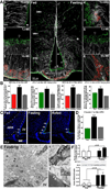
Fasting-induced fenestration of ME microvessel loops reaching the ARH, and tight junction complex reorganization in ARH tanycytes. (A) Vimentin (white), zonula occludens-1 (ZO-1, green) and MECA-32 (red) immunoreactivity in coronal sections of the hypothalamic tuberal region in fed and fasting mice. Tanycytic tight junction complexes exhibit a diffuse pattern (arrowheads, inset 1) when interacting with ZO-1-positive blood vessels (arrows, inset 1) and a honeycomb pattern (empty arrowheads, insets 2,4 and 5) when interacting with MECA-32-positive vessels (empty arrows, insets 3,4 and 6). (B) MECA-32-positive microvessel loop number and ZO-1-positive tight junction complex density in the ME and ARH according to nutritional status (n = 4 per group). (C) Distribution of claudin-1 (arrows, green) in coronal sections of the hypothalamic tuberal region in fed, fasting and refed mice. (D) Proportion of the ventricular wall facing the ARH immunolabeled for claudin-1 (n = 4 per group). (E) Electron micrographs from fasting mice showing ARH capillaries with nonfenestrated (arrow, inset 1) and fenestrated endothelia (arrowheads, inset 2); n: neuronal cell bodies; t: tanycyte cell bodies. Scale bar: 10 µm (1µm in insets). (F) Real-time PCR and immunoblotting for MECA-32 and actin from microdissected ME and MBH explants containing the ARH from fed and fasting mice (n = 3–4 per group). *** p<0.001; ** p<0.01; * p<0.05; fasting vs. fed and refed groups. DMH: dorsomedial nucleus of the hypothalamus.
Figure 2
Figure 2
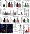
Fasting-induced BHB plasticity is mediated by glucose deprivation and tanycytic VEGF-A expression. (A) Blood glucose levels in fed and fasting mice (n = 6 per group). (B) Structural changes at the blood-CSF barrier in fasting mice infused with glucose (Glc; n = 6) or not (n = 6) and fed mice treated i.p. with 2-DG in the presence (n = 4) or absence of Axitinib (Axi; n = 4) compared to vehicle-treated fed mice (n = 3). (C) Real-time PCR analysis of genes involved in brain plasticity and glucosensing in the MBH of fasting and 2-DG-treated mice, normalized to values in mice fed ad libitum (red line) (n = 4 per group). (D) VEGF accumulates in the ME and MBH of fasting mice, as seen by immunoblotting (n = 3 per group). (E) 2-DG application to ME/vmARH explants increases VEGF secretion when compared to vehicle (red line) (n = 4 per group). (F) Quantification of MECA-32 and ZO-1 immunolabeling in fasting wild-type or VegfaloxP/loxP mice infused i.c.v. with Axitinib (n = 4) or tat-cre recombinant protein (n = 4), and fed mice treated with VEGF (n = 4) or vehicle (n = 10). (G) MECA-32 (red) and ZO-1 (green) immunolabeling in fasting mice and fed mice treated with VEGF or vehicle. Long MECA-32-positive microvessel loops reach the ependymal layer of the ME (arrows); arrowheads: MECA-32-positive intrainfundibular loop reaching the ARH. (H) Tanycyte isolation by FACS and real-time PCR analysis of VEGF-A mRNA in Tomato-positive (pos; tanycytes) and -negative cells (neg) in fed and fasting mice (n = 4 each). *** p<0.001; ** p<0.01; * p<0.05; various treatment groups vs. untreated fed mice.
Figure 3
Figure 3
Fasting-induced structural changes at the BHB facilitate the access of blood-borne metabolic signals to the ARH. (A) Evans Blue dye diffusion (gray) and MECA-32 (red) and ZO-1 (green) immunolabeling in the hypothalamic tuberal region in fed and fasting mice. (B) Quantification of Evans Blue diffusion into the ARH in fasting mice infused with Axitinib (n = 4) or glucose (Glc; n = 5), food-deprived tat-cre-treated VegfaloxP/loxP mice (n = 4), fed VEGF-treated mice (n = 5), food-deprived mice after refeeding for 24h (Refed; n = 4) and vehicle-treated fed mice (n = 7). (C) Placement of microdialysis cannulae (upper panel) for the simultaneous measurement of ARH and VMH glucose levels in fed and fasting rats, reported in the bar graph (lower panel, n = 4–5 per group). (D) Distribution of pSTAT3 immunoreactivity (white) in coronal sections of the ARH in fed and fasting mice treated or not with VEGF and Axitinib (Axi) after the i.p. administration of leptin (n = 4 per group), LAN (n = 3) or vehicle (n = 3–4 per group). (E) Quantification of pSTAT3 immunoreactive cells. (F) Structural differences between the ME and ARH of mice fed ad libitum and fasting mice, and their effects on the diffusion of blood-borne signals into the brain. *** p<0.001; ** p<0.01; * p<0.05; various treatment groups vs. untreated fed mice.
Figure 4
Figure 4
BHB plasticity modulates feeding behavior. (A) Cumulative and absolute food intake in fasting mice with or without i.p. Axitinib injection (orange; n = 4 per group) during refeeding after a 24h fast, and in vehicle-treated (gray; n = 4) or Axitinib-treated mice (gray and yellow stripes; n = 3–4) fed ad libitum. (B) Body weight gain during the first 48h of refeeding in mice previously food-deprived for 24h, and that were (orange; n =4) or not treated with Axitinib (black; n = 4). (C) Absolute food intake in mice fed ad libitum infused with VEGF (purple; n = 3) or not (gray; n = 4) for 3 days. (D) Anorectic and weight-loss-inducing effects of leptin and vehicle in mice fed ad libitum subjected to VEGF treatment or not (n = 10 per group). *** p<0.001; ** p<0.01; * p<0.05; various treatment groups vs. untreated fed mice.

Comment in

References

    1. Ambach G, Palkovits M. The blood supply of the hypothalamus of the rat. In: Morgane PJ, Panksepp J, editors. Handbook of the hypothalamus. New York: Marcel Dekker; 1979. pp. 267–377.
    1. Argaw AT, Gurfein BT, Zhang Y, Zameer A, John GR. VEGF-mediated disruption of endothelial CLN-5 promotes blood-brain barrier breakdown. Proc Natl Acad Sci U S A. 2009;106:1977–1982. - PMC - PubMed
    1. Banks WA. Blood-brain barrier and energy balance. Obesity (Silver Spring) 2006;14(Suppl 5):234S–237S. - PubMed
    1. Bouret SG, Bates SH, Chen S, Myers MG, Jr, Simerly RB. Distinct roles for specific leptin receptor signals in the development of hypothalamic feeding circuits. J Neurosci. 2012;32:1244–1252. - PMC - PubMed
    1. Carmeliet P, Dor Y, Herbert JM, Fukumura D, Brusselmans K, Dewerchin M, Neeman M, Bono F, Abramovitch R, Maxwell P, et al. Role of HIF-1alpha in hypoxia-mediated apoptosis, cell proliferation and tumour angiogenesis. Nature. 1998;394:485–490. - PubMed

Publication types

Substances