. 2013 Apr 18;38(4):681-93.
doi: 10.1016/j.immuni.2013.01.010. Epub 2013 Apr 4.

A network of high-mobility group box transcription factors programs innate interleukin-17 production

Collaborators, Affiliations

A network of high-mobility group box transcription factors programs innate interleukin-17 production

Nidhi Malhotra et al. Immunity. .

Abstract

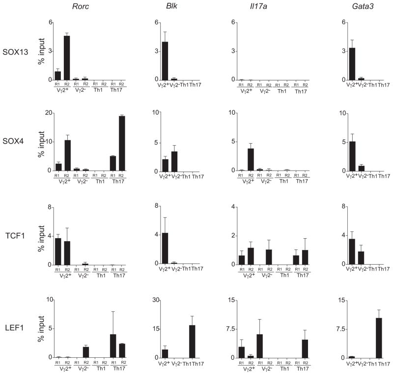
How innate lymphoid cells (ILCs) in the thymus and gut become specialized effectors is unclear. The prototypic innate-like γδ T cells (Tγδ17) are a major source of interleukin-17 (IL-17). We demonstrate that Tγδ17 cells are programmed by a gene regulatory network consisting of a quartet of high-mobility group (HMG) box transcription factors, SOX4, SOX13, TCF1, and LEF1, and not by conventional TCR signaling. SOX4 and SOX13 directly regulated the two requisite Tγδ17 cell-specific genes, Rorc and Blk, whereas TCF1 and LEF1 countered the SOX proteins and induced genes of alternate effector subsets. The T cell lineage specification factor TCF1 was also indispensable for the generation of IL-22 producing gut NKp46(+) ILCs and restrained cytokine production by lymphoid tissue inducer-like effectors. These results indicate that similar gene network architecture programs innate sources of IL-17, independent of anatomical origins.


Figures

Figure 1
Figure 1. SOX13 is essential for Tγδ17generation
(A) Frequencies of activated and mature Vγ2+ T cells in γδTCR+ cells in the spleen and thymus, respectively, of WT and Sox13−/− mice. Representative data (numbers within the gates represent percents of total) from one experiment of at least four are shown. Similar results were obtained with T-Sox13−/−mice (B6). (B) The defects in Tγδ17 generation originate in the thymus. LN and thymic mature (CD24lo) Vγ2+ cells from WT and Sox13−/− mice were analyzed for the expression of RORγt and EOMES (an activator of Ifng transcription), cell surface CCR6 and CD27 and intracellular IL-17A and IFNγ in matV2 cells. Frequencies less than 0.5% are left as blanks. (C) Intracellular staining for IL-17 in splenic V2 cells isolated from mice 4 hr post Zymosan administration. (D) Left, Intracellular and nuclear staining for the two markers of Tγδ17 cells, BLK and RORγt, in V2 thymocytes from neonatal mice at different maturational stages. Right, Staining of Abs to BLK and RORγt in CD4+ αβ thymocytes was used as negative controls. (E) SOX13 partly regulates RORγt expression in CD24hi immV2 thymocytes. A decrease in Rorc transcription (Top) as indicated by GFP expression from Rorc-Gfp substrate introduced to Sox13−/− mice, and intranuclear RORγt protein expression (Bottom). Representative data from one of two experiments is shown. (F) Intracellular staining for BLK in two maturation stages of Vγ2+ and Vγ2 γδ thymocytes from LCKp-Sox13 Tg mice. (G) Intracellular staining for IL-17A in Sox13 Tg+ LN γδ T cells. See also Fig. S1.
Figure 2
Figure 2. SOX4 regulates RORγt expression during Tγδ17 generation
(A) LN and mature thymic Vγ2+ cells from WT (CD2p-Cre:Sox4+/+) and T-Sox4−/− mice were analyzed for the expression of RORγt, CCR6 and CD27 and intracellular IL-17 and IFNγ. Representative data from one of four experiments is shown. (B) SOX4 regulates RORγt expression in immV2 thymocytes. The loss of Rorc transcription (Top) as indicated by the loss of GFP expression from Rorc-Gfp substrate introduced to T-Sox4−/− mice, and intranuclear RORγt protein expression (Bottom). Representative data from one of three experiments is shown. (C) Overlayed histograms of RORγt staining in αβ DP thymocytes in WT and T-Sox4−/− mice. The shaded histogram is the internal negative control for RORγt staining, gated on αβ CD4+ thymocytes that do not express Rorc. (D) PASI scoring was used to quantify the severity of psoriatic inflammation in IMQ treated mice. See also Fig. S2. Data is represented as mean+/− SEM.
Figure 3
Figure 3. TCF1 constrains Tγδ17generation
(A) Deregulated IL-17 production in Tcf7−/− mice. Differentiation of Tγδ17 thymocytes was examined by analyses of RORγt and EOMES, CCR6 and CD27, and intracellular IL-17A and IFNγ expression in mature (CD24lo) Vγ1.1+ and Vγ2+ γδ T cells. Similar results were obtained when peripheral γδ T cell subsets were analyzed. Representative profiles from one of at least five independent experiments, each with minimum of three mice/genotype, are shown. (B) Expression of CD27 expression on Tcf7−/− immV2 thymocytes. A similar trend for increased ratio of CCR6/CD27 was observed with other γδ thymic subtypes. (C) Left, Intranuclear staining for RORγt and LEF1 in LN Vγ1.1+ (top) and Vγ2+ (bottom) T cells from WT and Tcf7−/− mice shows mutually exclusive expression of the TFs and the loss of LEF1+ γδ T cells when TCF1 is non-functional. TCF1 expression, while biased, is not starkly separated from RORγt expressors in any γδ T cell subsets. Staining controls are shown in Figure S3E. (D) Intranuclear staining for LEF1 in immature (CD24hi) and mature (CD24lo) Vγ2+ thymocytes from WT and Tcf7−/− mice. See also Fig. S3.
Figure 4
Figure 4. ChIP assay for TF binding near the transcriptional regulatory sites of Rorc, Blk, il17a and Gata3 loci
Immature Vγ2+ and Vγ2 thymocytes, the immediate precursors of mature thymic effectors, were compared to in vitro differentiated control Th1 and Th17 CD4 αβ T cells. Analysis of mature γδ thymocytes was not possible due to their low numbers in mice. Graphs show quantitative PCR detection for relative enrichment of target DNA sequences from ChIP using Abs to indicated TF and control IgG (Fig. S4). The regions examined are described in Fig. S4 legend. Quantitative real-time PCR data are plotted as average percentage (%) of input +/−SD from two independent experiments. Binding of the TFs to TCF consensus sequences at the control MyoG promoter was undetectable in T cells (data not shown). See also Fig. S4.
Figure 5
Figure 5. Constrained impact of TCR signaling in effector specification
(A) PCA of the discriminatory gene signature of Itk−/− immV2 cells. PCA of the 15% most variable genes among the populations of cells shown (colors of bars and labels indicate population; MEV > 120 in at least one population; 1,433 genes). The first three principal components (PC1–PC3) are shown, along with the proportion of the total variability represented by each component (in parentheses along axes). (B) A heat map of relative gene expression of TFs in immature γδ subsets from WT and Itk−/− mice. Data were gene row normalized and hierarchically clustered by gene and subset. Genes are color coded (see legend) to display relative gene expression. (C) LN cells from WT and Tcrgv2 transgenic mice (with and without normal Sox13) that express a functional Vγ2-Jγ1-Cγ1 (Tcr Vg2 Tg) chain in nearly all γδ T cells (top) were analyzed for the expression of intracellular IL-17A in Vγ2+ T cells. Representative profiles from one of two experiments are shown, each with a minimum of three/group. Similar results were observed with thymocytes. (D) γδ T cell progenies of c-Kithi ETPs and c-Kit DN3 (CD25+CD44CD3CD4CD8) precursors cultured on OP9-DL1 stromal monolayers were assayed for CCR6 and CD27 expression. Representative FACS plots of V2 cells (top) and a summary of the frequencies (bottom, Student t-test P-values) of CCR6+CD27 Tγδ17 cells generated from ETPs (103 cells/well) or DN3 (5 ×10³ cells/well) precursors are shown. Similar results were obtained with varying cell numbers/well. Average cell numbers obtained from DN1 or DN3 were 3.5 × 10⁵ or 4.6 × 10⁴/well, respectively. V1/V6 = Vγ1.1+ cells. Data are combined from 3 independent experiments, ETP n=18; DN3 n=38. See also Fig. S5.
Figure 6
Figure 6. TCF1 regulates the differentiation and function of GALT ILCs
(A) TCF1/LEF1 expression in CD3CD19 mLN ILCs of adult mice. ILCs were segregated based on RORγt and IL-7R expression. CD4, NKp46, intranuclear TCF1 and LEF1 expression was assessed on the three indicated subsets. Data shown are representative profiles from one of three independent studies. (B) TCF1 expression in CD3CD19CD11b mLN ILCs segregated based on NKp46 and IL-7R was analyzed. (C) TCF1 is required for the development of NCR22 cells. Intestinal LP and splenic ILCs (CD3CD19IL-7R+) from Tcf7−/− neonates were stained with CD4 and NKp46 to track NCR22 cells. Data shown are representative profiles from one of four studies. (D) Frequencies of CCR6+ and CD25+ in neonatal LP ILCs from Tcf7+/− heterozygotes (HET) and Tcf7−/− mice. Lines represent the mean, and Student t-test P-values are shown. Similar results in mLN. (E) Numerical reduction in the ILC subsets in the mLN of 3 wk old Tcf7−/− mice. The total cell number of IL-7R+ ILCs, RORγthi and RORγtlo ILCs, and Nkp46+ and Nkp46 RORγtlo ILCs is shown. Data are combined from two experiments, n=5/group. Data is represented as mean+− SEM. (F) Representative histograms showing the frequency of NCR22 cells in the mLNs of 3 wk old Tcf7−/− mice. (G) Increased expression of RORγt in Tcf7−/− ILCs. Averages of MFI (+/−SEM) of RORγt expression in the CD3CD19IL-7Rhi spleen cells is provided (n=5/group; one representative experiment of four). (H) TCF1 restrains IL-17 and IL-22 production in the ILCs. Intracellular staining for IL-17 and IL-22 in the ex vivo splenic ILCs (CD3CD19IL-7RhiCD4+CD25+) was perfomed post-Zymosan administration. Unlike in other tissues, the number of splenic RORγt+ ILCs were marginally increased in adult Tcf7−/− mice. Profiles shown were obtained in two additional experiments. See also Fig. S6.

References

    1. Aliahmad P, de la Torre B, Kaye J. Shared dependence on the DNA-binding factor TOX for the development of lymphoid tissue-inducer cell and NK cell lineages. Nat Immunol. 2010;11:945–952. - PMC - PubMed
    1. Badis G, Berger MF, Philippakis AA, Talukder S, Gehrke AR, Jaeger SA, Chan ET, Metzler G, Vedenko A, Chen X, et al. Diversity and complexity in DNA recognition by transcription factors. Science. 2009;324:1720–1723. - PMC - PubMed
    1. Brustle A, Heink S, Huber M, Rosenplanter C, Stadelmann C, Yu P, Arpaia E, Mak TW, Kamradt T, Lohoff M. The development of inflammatory Th17 cells requires interferon-regulatory factor 4. Nat Immunol. 2007;8:958–966. - PubMed
    1. Buonocore S, Ahern PP, Uhlig HH, Ivanov II, Littman DR, Maloy KJ, Powrie F. Innate lymphoid cells drive IL-23-dependent innate intestinal pathology. Nature. 2010;464:1371–1375. - PMC - PubMed
    1. Cai Y, Shen X, Ding C, Qi C, Li K, Li X, Jala VR, Zhang HG, Wang T, Zheng J, Yan J. Pivotal role of dermal IL-17-producing γδT cells in skin inflammation. Immunity. 2011;35:596–610. - PMC - PubMed

Publication types

MeSH terms