. 2013 Jun;182(6):1986-95.
doi: 10.1016/j.ajpath.2013.02.008. Epub 2013 Apr 2.

Rac1 and Cdc42 differentially modulate cigarette smoke-induced airway cell migration through p120-catenin-dependent and -independent pathways

Affiliations

Rac1 and Cdc42 differentially modulate cigarette smoke-induced airway cell migration through p120-catenin-dependent and -independent pathways

Lili Zhang et al. Am J Pathol. 2013 Jun.

Abstract

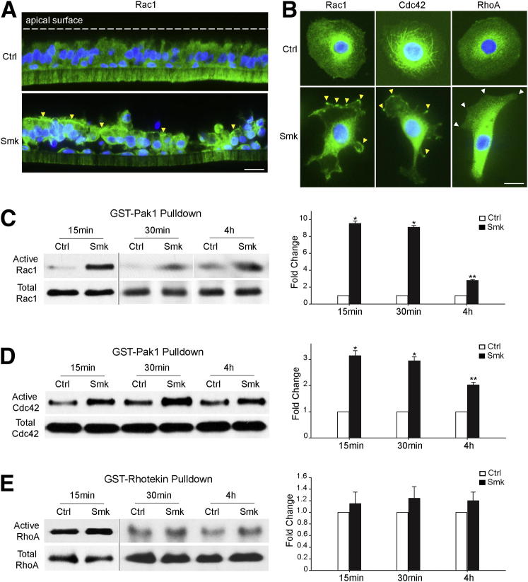
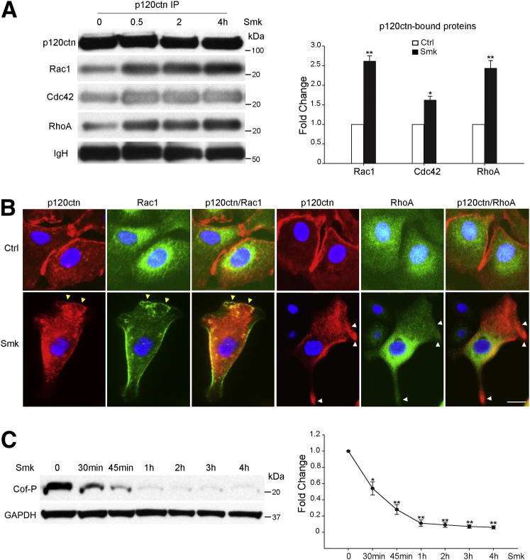
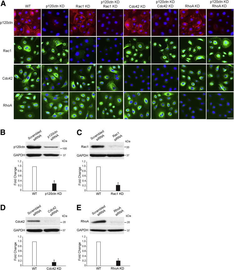
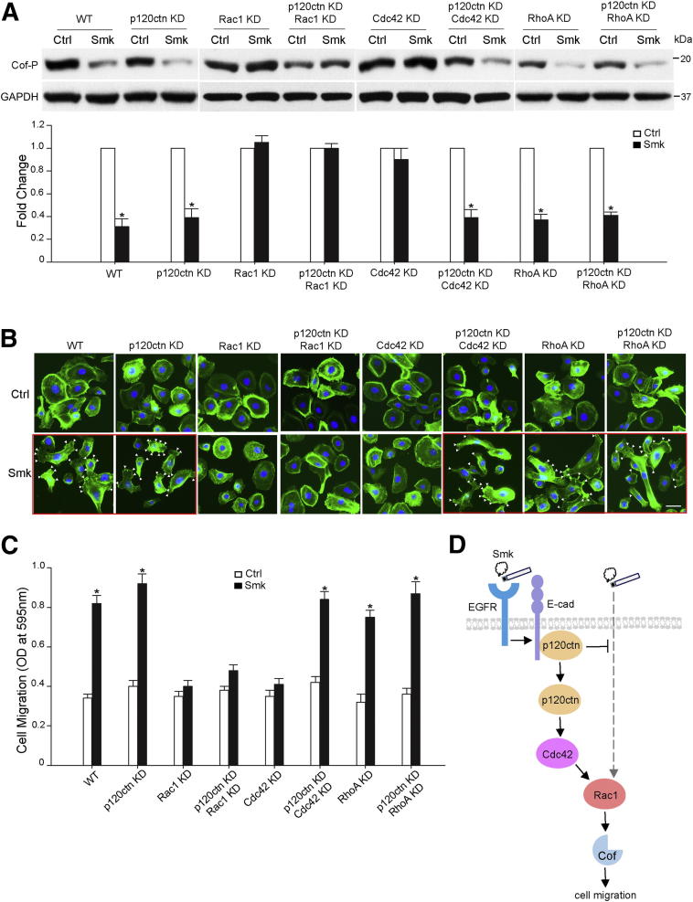
The adherens junction protein p120-catenin (p120ctn) shuttles between E-cadherin-bound and cytoplasmic pools to regulate E-cadherin/catenin complex stability and cell migration, respectively. When released from the adherens junction, p120ctn promotes cell migration through modulation of the Rho GTPases Rac1, Cdc42, and RhoA. Accordingly, the down-regulation and cytoplasmic mislocalization of p120ctn has been reported in all subtypes of lung cancers and is associated with grave prognosis. Previously, we reported that cigarette smoke induced cytoplasmic translocation of p120ctn and cell migration, but the underlying mechanism was unclear. Using primary human bronchial epithelial cells exposed to smoke-concentrated medium (Smk), we observed the translocation of Rac1 and Cdc42, but not RhoA, to the leading edge of polarized and migrating human bronchial epithelial cells. Rac1 and Cdc42 were robustly activated by smoke, whereas RhoA was inhibited. Accordingly, siRNA knockdown of Rac1 or Cdc42 completely abolished Smk-induced cell migration, whereas knockdown of RhoA had no effect. p120ctn/Rac1 double knockdown completely abolished Smk-induced cell migration, whereas p120ctn/Cdc42 double knockdown did not. These data suggested that Rac1 and Cdc42 coactivation was essential to smoke-promoted cell migration in the presence of p120ctn, whereas migration proceeded via Rac1 alone in the absence of p120ctn. Thus, Rac1 may provide an omnipotent therapeutic target in reversing cell migration during the early (intact p120ctn) and late (loss of p120ctn) stages of lung carcinogenesis.


Figures

Figure 1
Figure 1
Smoke activated Rac1 and Cdc42, but not RhoA, in migratory HBE cells. A: Rac1 (green) in pseudostratified HBE cells was stained by immunofluorescence after exposure to Ctrl and Smk for 4 hours. Nuclei were visualized with DAPI (blue). Yellow arrowheads point to apical areas where Rac1 accumulated in response to Smk exposure. B: Immunofluorescence staining of Rac1, Cdc42, and RhoA (all green) in HBE cells treated with Ctrl or Smk for 30 minutes. Nuclei were counterstained with DAPI (blue). Yellow arrowheads indicate localization of Rac1 and Cdc42 at the lamellipodia or filopodia of Smk-stimulated cells. White arrowheads indicate the absence of RhoA at the lamellipodia of Smk-treated cells. C–E: HBE cells were treated with Ctrl or Smk for the indicated times. Active Rac1 and Cdc42 were pulled down by a GST-Pak1 fusion protein and were analyzed by Western blot probed with anti-Rac1 (C) or anti-Cdc42 (D). Active RhoA was pulled down by a GST-rhotekin fusion protein and was detected by Western blot analysis with anti-RhoA (E). Total Rac1, Cdc42, and RhoA were used as loading controls. Relative fold change in active Rac1 (C), Cdc42 (D), or RhoA (E) after Smk treatment was normalized to untreated Ctrl (designated onefold), reported as means ± SEM fold change and graphed next to the representative blots. P < 0.01, ∗∗P < 0.05, Smk-treated cells versus Ctrl. Scale bars: 50 μm.
Figure 2
Figure 2
Smoke-mediated p120ctn/Rac1 and p120ctn/RhoA complex formation occurred in conjunction with smoke-induced dephosphorylation of cofilin. A: HBE cells were incubated with Ctrl or Smk for 0, 0.5, 2, and 4 hours. p120ctn immunoprecipitates (IPs) were immunoblotted for p120ctn, Rac1, Cdc42, and RhoA. Equal loading was revealed by immunoglobulin heavy chain (IgH). Densitometric quantitation of Rac1, Cdc42, and RhoA immunoprecipitated by p120ctn after 0.5 hours of Smk treatment was normalized to untreated Ctrl (designated onefold) and reported as means ± SEM fold change. B: HBE cells were incubated with Ctrl or Smk for 0.5 hours. Immunofluorescence staining of p120ctn (red), Rac1 (green), and RhoA (green) in HBE cells. Cell nuclei were counterstained with DAPI (blue). Yellow arrowheads indicate colocalization of p120ctn and Rac1 (merged yellow signals) at the leading edge of Smk-treated cells, and white arrowheads indicate the lack of p120ctn/RhoA colocalization in the lamellipodia and the leading edge of Smk-exposed cells. Scale bar = 50 μm. C: Cell lysates obtained from HBE cells treated with Ctrl or Smk for the indicated times were analyzed by Western blot with Cof-P. Equal loading was confirmed with glyceraldehyde-3-phosphate dehydrogenase (GAPDH). Relative fold change in Cof-P (reported as means ± SEM) induced by smoke was normalized to Ctrl (designated onefold) and graphed as a dose-response curve. P < 0.05, ∗∗P < 0.01.
Figure 3
Figure 3
siRNA knockdown of p120ctn, Rac1, Cdc42, and RhoA in primary HBE cells. HBE cells were transfected with scrambled siRNA (WT), p120ctn siRNA (p120ctn KD), Rac1 siRNA (Rac1 KD), p120ctn and Rac1 siRNA (p120ctn KD, Rac1 KD), Cdc42 siRNA (Cdc42 KD), p120ctn and Cdc42 siRNA (p120ctn KD, Cdc42 KD), RhoA siRNA (RhoA KD), and p120ctn and RhoA siRNA (p120ctn KD, RhoA KD), respectively. A: Cells were fixed 48 hours after transfection and were analyzed by immunofluorescence staining with p120ctn (red), Rac1 (green), Cdc42 (green), and RhoA (green). Cell nuclei were visualized with DAPI (blue). Scale bar = 50 μm. B–E: Cell lysates were obtained 48 hours after transfection and were analyzed by Western blot probed with p120ctn (B), Rac1 (C), Cdc42 (D), and RhoA (E). Glyceraldehyde-3-phosphate dehydrogenase (GAPDH) was used as loading control. Densitometric quantitation of protein levels in WT and KD cells 48 hours after transfection is graphed beneath the corresponding blot. The levels of proteins in KD cells were normalized to those in WT cells (designated onefold) and reported as means ± SEM fold change. P < 0.01, KD cells versus WT cells.
Figure 4
Figure 4
Rac1 mediated smoke-induced HBE cell migration in the presence and absence of p120ctn, whereas Cdc42 functioned only in the presence of p120ctn. A–C: HBE cells were transfected with scrambled siRNA (WT), p120ctn siRNA (p120ctn KD), Rac1 siRNA (Rac1 KD), p120ctn and Rac1 siRNA (p120ctn KD, Rac1 KD), Cdc42 siRNA (Cdc42 KD), p120ctn and Cdc42 siRNA (p120ctn KD, Cdc42 KD), RhoA siRNA (RhoA KD), and p120ctn and RhoA siRNA (p120ctn KD, RhoA KD), respectively. A and B: Forty-eight hours after transfection, HBE cells were treated with Ctrl and Smk for 4 hours. A: Cell lysates were obtained and analyzed by Western blot with Cof-P. Equal loading was confirmed with glyceraldehyde-3-phosphate dehydrogenase (GAPDH). Relative fold change of Cof-P in Smk-exposed cells was normalized to unexposed Ctrl (designated onefold) and reported as means ± SEM fold change. B: HBE cells were fixed and stained with fluorescein isothiocyanate–labeled phalloidin to reveal actin (green). Cell nuclei were counterstained with DAPI (blue). Frames with the red outline indicate treatment conditions under which HBE cell migration was preserved. White arrowheads indicate the formation of lamellipodia in Smk-stimulated cells. Scale bar = 50 μm. C: Forty-eight hours after transfection, siRNA-transfected cells were loaded on Boyden chambers and incubated with Ctrl or Smk for 4 hours. Migrating cells on the bottom side of the membrane were quantified by measuring the OD at 595 nm and plotted as the means ± SD of three independent chambers. D: Differential role of Rac1 and Cdc42 in regulating smoke-promoted HBE cell migration. In the normal human airway, the AJ proteins E-cad and p120ctn mediate cell-cell adhesion. Smoke disrupts AJs and provokes the cytoplasmic translocation of p120ctn through EGFR signaling., , In the early stages of lung cancer, when intracellular p120ctn is abundant, smoke promotes the activation (dephosphorylation) of actin-severing protein, Cof, to promote cell migration. Rac1 and Cdc42 are implicated in this p120ctn-dependent pathway because knocking down either Rac1 or Cdc42 abrogated smoke-induced cell migration in the presence of p120ctn. In the advanced stages of lung cancer, when p120ctn is lost, Cof dephosphorylation is mediated through Rac1. The alternative Rac1 pathway seems to be suppressed by membrane p120ctn and only unveiled after the loss of tumor-suppressing p120ctn. The Rac1 pathway differs from the p120ctn/Rac1 pathway in that its action does not require Cdc42. Thereby, knockdown of Rac1 alone succeeds in abolishing smoke-induced cell migration in the absence of p120ctn. Because Rac1 activation is essential for the p120ctn-dependent and p120ctn-independent cell migratory pathways induced by smoke, Rac1 likely acts at the convergent point of the two pathways and, thus, downstream of Cdc42. The present data support the potential of Rac1 as an omnipotent therapeutic target in treating the early and advanced stages of lung cancer. P < 0.01, Smk-treated cells versus Ctrl.

Similar articles

Cited by

References

    1. IARC Working Group on the Evaluation of Carcinogenic Risks to Humans Tobacco smoke and involuntary smoking. IARC MonogrEval Carcinog Risks Hum. 2004;83:1–1438. - PMC - PubMed
    1. Proctor R.N. The global smoking epidemic: a history and status report. Clin Lung Cancer. 2004;5:371–376. - PubMed
    1. Hecht S.S., Kassie F., Hatsukami D.K. Chemoprevention of lung carcinogenesis in addicted smokers and ex-smokers. Nat Rev Cancer. 2009;9:476–488. - PMC - PubMed
    1. Dasari V., Gallup M., Lemjabbar H., Maltseva I., McNamara N. Epithelial-mesenchymal transition in lung cancer: is tobacco the “smoking gun”? Am J Respir Cell Mol Biol. 2006;35:3–9. - PubMed
    1. Gomperts B.N., Spira A., Massion P.P., Walser T.C., Wistuba I.I., Minna J.D., Dubinett S.M. Evolving concepts in lung carcinogenesis. Semin Respir Crit Care Med. 2011;32:32–43. - PMC - PubMed

Publication types

MeSH terms