. 2013 Jul 5;441(2):95-106.
doi: 10.1016/j.virol.2013.03.014. Epub 2013 Apr 4.

Metagenomic study of the viruses of African straw-coloured fruit bats: detection of a chiropteran poxvirus and isolation of a novel adenovirus

Affiliations

Metagenomic study of the viruses of African straw-coloured fruit bats: detection of a chiropteran poxvirus and isolation of a novel adenovirus

Kate S Baker et al. Virology. .

Abstract

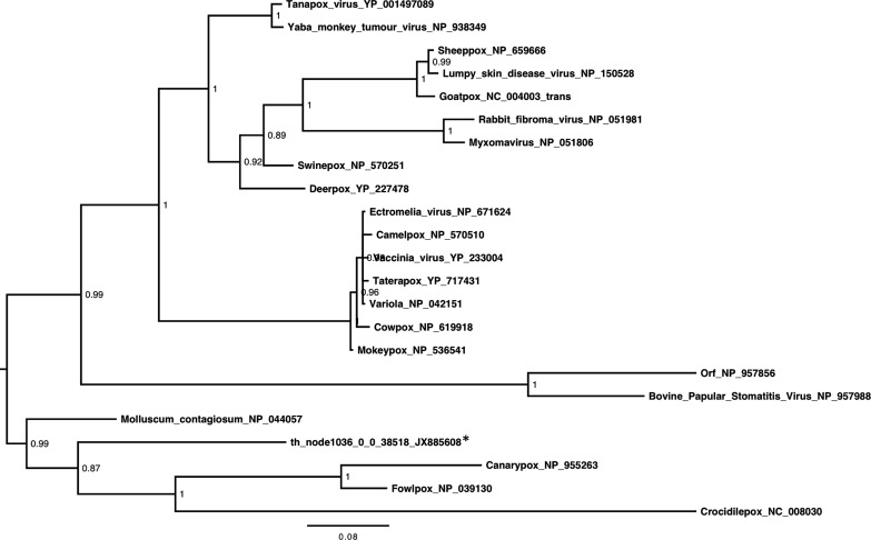
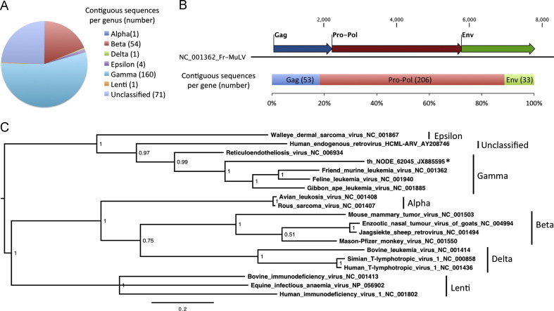
Viral emergence as a result of zoonotic transmission constitutes a continuous public health threat. Emerging viruses such as SARS coronavirus, hantaviruses and henipaviruses have wildlife reservoirs. Characterising the viruses of candidate reservoir species in geographical hot spots for viral emergence is a sensible approach to develop tools to predict, prevent, or contain emergence events. Here, we explore the viruses of Eidolon helvum, an Old World fruit bat species widely distributed in Africa that lives in close proximity to humans. We identified a great abundance and diversity of novel herpes and papillomaviruses, described the isolation of a novel adenovirus, and detected, for the first time, sequences of a chiropteran poxvirus closely related with Molluscum contagiosum. In sum, E. helvum display a wide variety of mammalian viruses, some of them genetically similar to known human pathogens, highlighting the possibility of zoonotic transmission.


Figures

Fig. 1
Fig. 1
Bioinformatic analysis pipeline for 15,155,461 assembled contigs. (A) Contigs assembled from sequencing reads by different de novo assemblers (starred, denoted by colours) were consolidated by sequential comparisons (numbered, curved green arrows) and removal (red arrows) of duplicate sequences. The size of charts is proportional to the number of contigs. (B) Consolidated sequences subject to sequential BLAST comparison with automated taxonomic classification to identify suspect-viral sequences. Then sequences were excluded manually on the basis of related viral family and curation to identify a final set of viral sequences. Proportions of sequences assembled by each algorithm (coloured as in A) and from each sample type (coloured as in the inset key) before and after length-exclusion are shown in the stacked charts. (C) Proportions of 1,363 mammalian-virus related contigs by assembly algorithm, sample-type and identification algorithm (shaded as in B) are shown in the stacked chart. Proportions by viral family are shown in the chart on the right. Number of contigs related to members of each family are shown in parentheses after the viral family names, which are grouped by genome type. (For interpretation of the references to color in this figure legend, the reader is referred to the web version of this article.)
Fig. 2
Fig. 2
Presence of diverse herpesviruses in throat swab samples fromEidolon helvum. Midpoint-rooted phylogenetic tree based on 166aa of the DNA polymerase protein encoded by three contigs (starred) and representatives from the Herpesviridae. Herpesvirinae subfamily clusters are shown. Posterior probability values are shown and the bar represents the expected number of amino acid substitutions per site.
Fig. 3
Fig. 3
Gene origin and diversity of papillomavirus sequences present inE. helvum. (A) Top: schematic representation of Rousettus aegyptiacus papillomavirus 1 (RaPV-1) genome. Bottom: gene classification of 408 contigs identified as papillomaviruses. The pie chart shows the proportion of contigs related to each gene. The number of contigs is shown in brackets after the gene name. (B) Midpoint-rooted phylogenetic tree based on 157 aa of the major capsid (L1) protein encoded by six contigs (starred) and representatives from the Papillomaviridae. Posterior probability values are shown at each node and the bar represents the expected number of amino acid substitutions per site.
Fig. 4
Fig. 4
Isolation ofE. helvumadenovirus 1 (AdV1). (A) Pteropus alecto primary kidney cells showing cytopathic effect (and control) at day 6 post infection with E. helvum AdV1. Magnification is 200 X (B) Transmission electron micrograph of a Pteropus alecto cell infected with E. helvum AdV1. Arrow indicates adenovirus particles observed in the nucleus (N). Bar represents 200 nm. Insert adenovirus particle negative stained from supernatant of the same culture. Insert bar represents 100 nm. (C) Midpoint-rooted phylogenetic tree based on alignment of hexon proteins (915aa) of E. helvum AdV1 (starred) and representative adenoviruses. Genera are demarcated by adjacent lines and names. Posterior probability values are shown and the bar represents the expected number of amino acid substitutions per site.
Fig. 5
Fig. 5
Phylogenetic analysis of anE. helvumpoxviral sequence. Midpoint-rooted phylogenetic tree based on 286 aa of the major core protein encoded by a contig from the throat sample (starred) and representatives from the Poxviridae. Posterior probability values are shown and the bar represents the expected number of amino acid substitutions per site.
Fig. 6
Fig. 6
Classification and phylogenetic analysis of retroviral sequences present inE. helvum. (A) The chart shows proportions of retroviral sequences related to different retroviral genera. The number of sequences related to each genus is shown in parentheses. (B) Retroviral sequences classified by the gene they encode. The top bar represents the genome organization of Friend-murine leukemia virus, with numbers representing genome nucleotide positions. The lower bar shows the proportion of 292 retroviral contigs that were related to each gene. The number of sequences related to each gene is shown in parentheses. (C) Midpoint-rooted phylogenetic tree based on 176aa of the Pro-Pol gene of representative retroviruses and a contig from the throat sample (starred), posterior probability values are shown at each node and the bar represents the expected number of amino acid substitutions per site.

References

    1. Allander T., Emerson S.U., Engle R.E., Purcell R.H., Bukh J. A virus discovery method incorporating DNase treatment and its application to the identification of two bovine parvovirus species. PNAS. 2001;98:11609–11614. - PMC - PubMed
    1. Altschul S.F., Gish W., Miller W., Myers E.W., Lipman D.J. Basic local alignment search tool. J. Mol. Biol. 1990;215:403–410. - PubMed
    1. Antia R., Regoes R.R., Koella J.C., Bergstrom C.T. The role of evolution in the emergence of infectious diseases. Nature. 2003;426:658–661. - PMC - PubMed
    1. Baker K.S., Todd S., Marsh G., Fernandez-Loras A., Suu-Ire R., Wood J.L., Wang L.F., Murcia P.R., Cunningham A.A. Co-circulation of diverse paramyxoviruses in an urban African fruit bat population. J. Gen. Virol. 2012;93:850–856. - PMC - PubMed
    1. Bermingham, A., Chand, M.A., Brown, C.S., Aarons, E., Tong, C., Langrish, C., Hoschler, K., Brown, K., Galiano, M., Myers, R., Pebody, R.G., Green, H.J., Boddington, N.L., Gopal, R., Price, N., Newsholme, W., Drosten, C., Fouchier, R.A., Zambon, M., 2012. Severe Respiratory Illness Caused by a Novel Coronavirus, in a Patient Transferred to the United Kingdom from the Middle East, September 2012. Eurosurveillance 17, Available online: 〈http://www.eurosurveillance.org/ViewArticle.aspx?ArticleId=20290〉. - PubMed

Publication types