. 2013 Jun 1;378(1):1-12.
doi: 10.1016/j.ydbio.2013.02.024. Epub 2013 Apr 3.

Different thresholds of Wnt-Frizzled 7 signaling coordinate proliferation, morphogenesis and fate of endoderm progenitor cells

Affiliations

Different thresholds of Wnt-Frizzled 7 signaling coordinate proliferation, morphogenesis and fate of endoderm progenitor cells

Zheng Zhang et al. Dev Biol. .

Abstract

Wnt signaling has multiple dynamic roles during development of the gastrointestinal and respiratory systems. Differential Wnt signaling is thought to be a critical step in Xenopus endoderm patterning such that during late gastrula and early somite stages of embryogenesis, Wnt activity must be suppressed in the anterior to allow the specification of foregut progenitors. However, the foregut endoderm also expresses the Wnt-receptor Frizzled 7 (Fzd7) as well as several Wnt ligands suggesting that the current model may be too simple. In this study, we show that Fzd7 is required to transduce a low level of Wnt signaling that is essential to maintain foregut progenitors. Foregut-specific Fzd7-depletion from the Xenopus foregut resulted in liver and pancreas agenesis. Fzd7-depleted embryos failed to maintain the foregut progenitor marker hhex and exhibited decreased proliferation; in addition the foregut cells were enlarged with a randomized orientation. We show that in the foregut Fzd7 signals via both the Wnt/β-catenin and Wnt/JNK pathways and that different thresholds of Wnt-Fzd7 activity coordinate progenitor cell fate, proliferation and morphogenesis.


Figures

Figure 1
Figure 1. Differential Wnt signaling patterns the Xenopus endoderm
Wnt signaling has different thresholds in the endoderm. Embryos were injected with a dose range of mRNA encoding Sfrp5; 500 pg (B,G), 800 pg (C,H), 2 ng (D,I) and 3 ng (E,J). In situ hybridization for hhex (A–E) and vent1 and vent2, referred to as vent1/2 (F–J) in stage 19 bisected embryos showed that low doses of Sfrp5 expanded the hhex expression domain (yellow dashed line) (B–D) at the expense of hindgut markers vent1 and vent2 (a mixture of both probes referred to as vent1/2) (G–I). The highest dose of Sfrp5 resulted in a loss of hhex (E). The number of embryos with the illustrated phenotype is indicated in each panel.
Figure 2
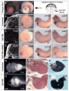
Figure 2. Fzd7-depletion disrupts foregut organogenesis
(A) Targeted injection of fzd7-MOs (50 ng) and red fluorescent tracer (RLDx) into the D1 cells of 32-cell stage Xenopus embryos. Bight field (BF) and fluorescent view of representative bisected stage 20 embryo showing that the injection targeted the foregut (fg) and avoided the axial mesoderm. (B–D) Confocal immunostaining confirmed that the fzd7-MOs resulted in a loss of Fzd7 protein from the foregut cell membrane (B,C), which was rescued by injection of Fzd7 mRNA lacking MO-target sequence (D). (E–M) In situ hybridization showed that fzd7-MO embryos failed to maintain hhex at stage 20 and did not express the liver (nr1h5) and pancreas/duodenum (pdx1) markers at stage 35, which could be partially rescued by injection of Fzd7 mRNA. (N, O) Ventral view of stage 45 embryos showing foregut edema and defective intestinal coiling with shortened gut in fzd7-MO injected embryos. (P,Q) H&E-stained section of a control (P) and a fzd7-MO-injected embryo (Q) that lacks foregut organs including liver (lv) and stomach (st). (R, S) In situ hybridization of a2m in isolated stage 42 gut tubes showed loss of foregut organs such as liver (lv), stomach (st) and pancreas (p) and shortened intestine (In). The number of embryos with the illustrated phenotype is indicated in each panel.
Figure 3
Figure 3. Fzd7-depletion causes defects in foregut cell morphology
(A–B) Bight field view of the foregut surface at stage 20 showed that Fzd7 morphants exhibit larger loosely adherent cells (B), compared to controls (A). (C–J) Confocal immunostaining of the foregut (anterior left, dorsal up) with μ1-integrin (C,D), C-cadherin (E,F), β-catenin (G,H) and phalloidin (F-actin) (I,J) showed decreased cell adhesion molecules and reduced cytoskeleton in the enlarged foregut cells of Fzd7 morphants. All of the images were taken using same setting for control and fzd7-MO embryos. (K–L) Quantitation of cell size and orientation; foregut cell length, width and orientation in control (K) and fzd7-MO injected embryos (L) were measured from β-catenin immunostaining (green) using Image-J. Nuclei shown in blue, fzd7-MO/RLDx in red. All images were oriented with dorsal up. The yellow line marks the long axis of foregut cells, and quantification shows the frequency of orientations of the long axis of cells in Fzd7 morphants (L,L′) and control (K,K′) n= the total number of foregut cells from 5 uninjected (K′) and 5 fzd7-MO embryos (L′). (M) The relative length, width, and length/width radio in control and Fzd7-depleted foregut cells. **p<0.01 relative to controls in Student’s t-test (N) Western blot analysis shows no significant changes of total E-cadherin and C-cadherin level in the foregut explants.
Figure 4
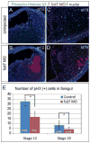
Figure 4. Fzd7-depletion causes reduced cell proliferation
(A–D) Confocal immunostaining of phospho-Histone h3 (PH3; green), nuclei (blue) and fzd7-MO/RLDx (red) at stage 12 (A,B) and stage 19 (C,D) show that Fzd7-depleted embryos exhibit reduced foregut (outlined in dashed yellow line) proliferation. (E) Mean number of PH3 positive cells in the foregut +/− S.D. *p<0.05 relative to sibling controls in Student’s t-test (n= 4 embryos/condition).
Figure 5
Figure 5. Fzd7 depletion results loss of both Wnt/β-catenin and Wnt/JNK activity in the foregut
(A) Fzd7-depletion resulted in a reduction of β-catenin/Tcf and JNK/AP1 activity in the foregut. TOP:flash or AP1:Luciferase reporter plasmids were injected into either the D1 foregut endoderm cells or the D4 hindgut endoderm cells at the 32-cell stage, with or without fzd7-MO as indicated. The TOP:Flash reporter is an indicator of β-catenin/Tcf activity, while the AP1:luciferase reporter is an indicator of JNK-mediated c-Jun/c-Fos (AP1) activity. At stage 20 luciferase activity was measured, in triplicate. The average relative luciferase activity, normalized to co-injected pRTK:Renila, from three biological replicates per condition is shown +/− S.D. *p<0.05 and ** p<0.01 relative to control foregut in Student’s t-test. (B) Western blot showed decreased phospho-JNK1/2 (p-JNK) and a loss of dephosphorylated active β-catenin and total cytosolic β-catenin in the foregut explants at stage 19. (C–D): Confocal immunostaining showed reduced nuclear β-catenin levels in Fzd7 morphant foregut tissue (D) relative to controls (C), at stage 20. (E) Mean pixel intensity of nuclear β-catenin staining measured using Image-J +/− S.D (foregut cells were scored from 5 embryos/condition).
Figure 6
Figure 6. Fzd7 signals via both β-catenin and JNK coordinate foregut cell proliferation, gene expression and cell morphology
(A) Inhibition of either Wnt/β-catenin or JNK pathways reduced foregut cell proliferation. Embryos were either injected with RNA encoding Dkk1 (500 pg) to block the Wnt/β-catenin pathway or treated with the cell soluble JNK inhibitor SB600125 (JNKi; 100 μM). Mean number of PH3 positive cells in the foregut +/− S.D. *p<0.05 and ** p<0.01 compared to controls (n=4 embryos/condition). (B) Activation of either β-catenin or JNK signaling rescued cell proliferation in Fzd7 morphants. Embryos were injected with fzd7-MOs (50 ng) with or without RNA encoding a constitutively active JNK (c.a.JNK; 200 pg) or a hormone inducible GR:Lef-βCTA (β-cat; 200 pg) activated by 1 μM dexamethasone at stage 11. Mean number of P-H3 positive foregut cells at stage 12 +/− S.D. *p<0.05 relative to control and **p<0.05 relative to fzd7-MO alone in Student’s t-test (n= 4 embryos/condition). (C–H) Fzd7/JNK signaling regulates cell shape. Confocal immunostaining of β-catenin at stage 20 showed that c.a.JNK injection (F) rescued the cell-size defects in Fzd7 morphants (D), whereas activation of the GR:Lef-βCTA (β-cat) did not (E). The JNK inhibitor (JNKi) caused a reduction of cytoskeletal β-catenin and increased cell size (H), phenocopying fzd7-MO (D), whereas Dkk1 (1.5 ng) had no effects on cell morphology (G). (I–N) Both Fzd7/β-catenin and Fzd7/JNK regulate gene expression. In situ hybridization to stage 20 embryos showed that co-injection of either GR:Lef-βCTA (β-cat) (K) or c.a.JNK (L) restored hhex expression in Fzd7 morphants (J), whereas the JNK inhibitor (N) or high levels of Dkk1 (1.5 ng) (M) suppressed hhex. The number of embryos with the illustrated phenotype is indicated in each panel.
Figure 7
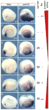
Figure 7. Multiple thresholds of Wnt/Fzd7/β-catenin activity pattern the endoderm
(AL) Fzd7/β-catenin signaling levels were modulated in a dose response with different combination of fzd7-MOs and/or treatment with BIO, a GSK3 inhibitor that stabilizes β-catenin. In situ hybridization of the foregut marker hhex and the mid/hindgut markers vent1/2 in stage 20 embryos showed that increasing dose of BIO and therefore increasing β-catenin activity decreased hhex while expanding vent1/2 (A, B) relative to untreated controls (C, D). A partial knockdown of Fzd7 (25 ng of fzd7-MOs) resulted in a modest increase in hhex and reduction of vent1/2 (E, F), whereas a complete Fzd7 knockdown by 50 ng of the fzd7-MO caused a loss of hhex, which was not accompanied by an expansion of vent1/2 (G, H). A low dose of BIO (5 uM in I,J) rescued hhex expression in fzd7-depleted embryos (I, J), whereas a higher BIO dose (10 uM) resulted in the foregut adopting a hindgut fate and expressing ectopic vent1/2 (K, L). The number of embryos with the illustrated phenotype is indicated in each panel.
Figure 8
Figure 8. A model of how Wnt/Fzd7/Sfrp5 regulate β-catenin and JNK signaling to coordinate endoderm fate, morphogenesis
(A) Schematic of a neurula embryo (anterior left) showing expression of the fzd7 (green), wnt11, wnt5 and wnt8 in the ventral mesoderm and endoderm (blue) and the Wnt-antagonist sfrp5 in the surface of the foregut endoderm (red). The spatial expression pattern of receptors, ligands and antagonists is postulated to establish differential Wnt activity in the endoderm. (B) Different thresholds of β-catenin/TCF activity (red line) pattern endoderm with high activity promoting hindgut progenitor fate (hg), where as a low but essential level of β-catenin/TCF is required to maintain foregut (fg) fate. (C) Differential Fzd7/JNK activity might regulate cell shape, adhesion and morphogenesis in the foregut (green). Fzd7/JNK signaling in the deep endoderm promotes cell adhesion and the oriented cell shape required for gut elongation. Sfrp5 in the surface layer reduces JNK activity to a low but essential level to establish an epithelium. If JNK activity is too low (as in Fzd7 morphants) cells become enlarged, loosely adherent with a random orientation.

Similar articles

Cited by

References

    1. Adams CL, Nelson WJ, Smith SJ. Quantitative analysis of cadherin-catenin-actin reorganization during development of cell-cell adhesion. J Cell Biol. 1996;135:1899–1911. - PMC - PubMed
    1. Bovolenta P, Rodriguez J, Esteve P. Frizzled/RYK mediated signalling in axon guidance. Development. 2006;133:4399–4408. - PubMed
    1. Brown JD, Hallagan SE, McGrew LL, Miller JR, Moon RT. The maternal Xenopus beta-catenin signaling pathway, activated by frizzled homologs, induces goosecoid in a cell non-autonomous manner. Development, growth & differentiation. 2000;42:347–357. - PubMed
    1. Cha SW, Tadjuidje E, Tao Q, Wylie C, Heasman J. Wnt5a and Wnt11 interact in a maternal Dkk1-regulated fashion to activate both canonical and non-canonical signaling in Xenopus axis formation. Development. 2008;135:3719–3729. - PubMed
    1. Cha SW, Tadjuidje E, White J, Wells J, Mayhew C, Wylie C, Heasman J. Wnt11/5a complex formation caused by tyrosine sulfation increases canonical signaling activity. Curr Biol. 2009;19:1573–1580. - PubMed

Publication types