. 2013 Apr 11;153(2):438-48.
doi: 10.1016/j.cell.2013.03.006. Epub 2013 Apr 4.

The bacterial DnaC helicase loader is a DnaB ring breaker

Affiliations

The bacterial DnaC helicase loader is a DnaB ring breaker

Ernesto Arias-Palomo et al. Cell. .

Abstract

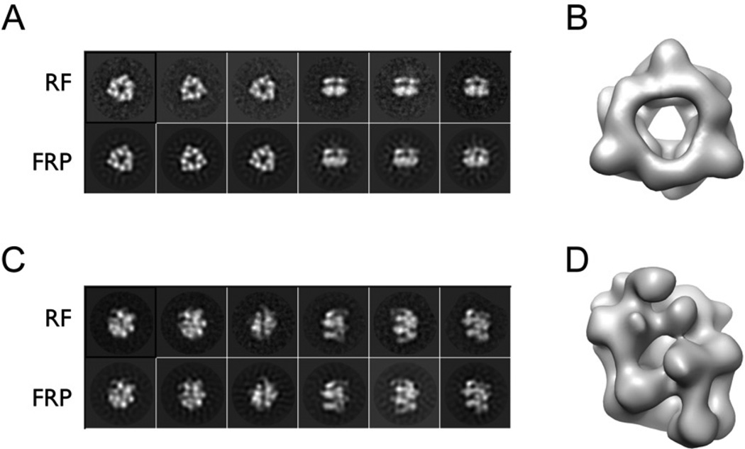
Dedicated AAA+ ATPases deposit hexameric ring-shaped helicases onto DNA to promote replication in cellular organisms. To understand how loading occurs, we used electron microscopy and small angle X-ray scattering (SAXS) to determine the ATP-bound structure of the intact E. coli DnaB⋅DnaC helicase/loader complex. The 480 kDa dodecamer forms a three-tiered assembly, in which DnaC adopts a spiral configuration that remodels N-terminal scaffolding and C-terminal motor regions of DnaB to produce a clear break in the helicase ring. Surprisingly, DnaC's AAA+ fold is dispensable for ring remodeling because the DnaC isolated helicase-binding domain can both load DnaB onto DNA and increase the efficiency by which the helicase acts on substrates in vitro. Our data demonstrate that DnaC opens DnaB by a mechanism akin to that of polymerase clamp loaders and indicate that bacterial replicative helicases, like their eukaryotic counterparts, possess autoregulatory elements that influence how hexameric motor domains are loaded onto and unwind DNA.


Figures

Figure 1
Figure 1. EM reconstructions of E.coli DnaB and DnaBC
(A) Representative reference-free 2D class averages of DnaB (RF) compared to forward reprojections of the 3D structure obtained after multi-reference refinement (FRP). (B) The 3D reconstruction of DnaB shows a closed ring with a broad inner channel. (C) Representative reference-free 2D class averages of the DnaBC complex (RF) compared to forward reprojections of the 3D structure obtained after multi-reference refinement (FRP). (D) The 3D reconstruction for DnaBC shows a 3-tiered, cracked-ring particle. See Figures S1–S4.
Figure 2
Figure 2. EM analysis of E.coli DnaB and DnaBC
(A) Closed-ring B. stearothermophilus DnaB crystal structure (PDB ID 2R6A, (Bailey et al., 2007b)) fitted in the E.coli DnaB EM reconstruction. (B) Closed-ring B. stearothermophilus DnaB crystal structure (PDB ID 2R6A) filtered at 25 Å. (C) DnaBC forms a three-tiered, right-handed, cracked-ring structure. The upper tier is colored in blue (1), middle in purple (2) and lower in orange (3). (D) DnaBC view showing the discontinuity in the cracked ring. The orientation is orthogonal to that of panel C. (E) The DnaBC lower tier exhibits 61 symmetry. (F) The DnaBC upper tier exhibits 31 symmetry. (G) Longitudinal DnaBC section, showing the central channel that runs along the three tiers. The orientation is equivalent to that of panel D (H) DnaBC displayed at lower threshold (5.7 σ contour) to highlight the discontinuity of the ring. This view is equivalent to that of panel D.
Figure 3
Figure 3. Fitting DnaB into the DnaBC model
(A) Primary structure of E.coli DnaB and E.coli DnaC. (B) Structure of a typical DnaB N-terminal domain homodimer (PDB ID 2R6A, (Bailey et al., 2007b)). (C) Docking of DnaB N-terminal domain homodimers into tier 1 of the DnaBC model. Subunits are alternatingly colored as per panel B. (D) Docking of DnaB RecA folds into tier 2 of the DnaBC model. Subunits are alternatingly colored purple and pink. The density corresponding to the third tier has been removed for clarity. (E) Side view of both domains of DnaB fitted into the DnaBC complex. The density corresponding to the third tier is not shown.
Figure 4
Figure 4. Molecular architecture of the DnaBC complex
(A) Detail view of DnaCAAA+ crystal structure (PDB ID 3ECC (Mott et al., 2008)). The DnaC ATPase domains appear to productively associate with nucleotide binding in this complex. (B) DnaCAAA+ hexamer (reconstituted by imposing the crystallographically-observed 61 packing arrangement on PDB ID 3ECC), fitted into the lower tier of the DnaBC complex. (C) Composite model for DnaBC. The six columns of unaccounted density connecting the DnaCAAA+ and DnaB RecA domains likely correspond to the DnaC N-terminal regions, which bind DnaB. See Movie S1. (D) SAXS analysis of DnaBC. Experimental scattering data (1 mM AMPPNP, pink) fits well to a theoretical curve calculated from the DnaBC EM reconstruction (blue), but not to that of a closed-ring DnaB hexamer (purple). Improved fitting is obtained from a multiple ensemble search containing a mixture of DnaBC model and free DnaB (green). See Figure S2. (E) Clamp-loader AAA+ domains oligomerize into a right-handed spiral to open a processivity clamp (PDB ID 3U5Z (Kelch et al., 2011)).
Figure 5
Figure 5. The DnaB N-terminal domain collar is remodeled by DnaC
(A) The N-terminal homodimers of DnaB in the absence of nucleotide form a wide, closed triangular collar (PDB ID 2R6A, (Bailey et al., 2007b)). DnaB RecA domains are displayed as surfaces, whereas the N-terminal domains and linker helices are shown as light blue/orange cylinders. (B) The N-terminal domains of DnaB within the DnaBC complex undergo a marked positional shift from the closed ring state, forming new packing arrangements between dimers. The DnaB RecA domains form a cracked spiral (boxed inset); DnaC is omitted for clarity. A schematic of the arrangement for the N-terminal domain dimers is shown in the upper left corner of each panel. Arrows in panel A indicate the movement of the DnaB NTDs to the state shown in panel B. See Figures S5 and S6 and Movies S2 and S3.
Figure 6
Figure 6. Intrinsic and extrinsic control of DnaB ring opening and function
(A) Removal of the N-terminal domain of the DnaB-family G40P protein allows its associated RecA ATPase to assemble into a helical filament (PDB ID 3BH0, (Wang et al., 2008)). (B) The fitted DnaB RecA domains within DnaBC structure adopt a spiral pitch similar to that seen for N-terminally truncated G40P. (C) The DnaC NTD can promote the DnaB-dependent unwinding of a topologically-closed DNA substrate that requires helicase ring opening and loading. (Top) Gel showing unwinding of a 3’ tailed oligonucleotide annealed to circular M13mp18 ssDNA in the presence of DnaB with or without DnaC or the DnaC NTD. Lanes N and D indicate the native and boiled substrate, respectively. (Bottom) Quantification of product seen by gel. Concentrations of DnaB hexamers in the reactions are shown; DnaC, when present, was included at a 2-fold molar excess of DnaB monomers. Columns and error bars represent the average and standard deviations, respectively, of at least five measurements. (D) DnaC and the DnaC NTD can promote the DnaB-dependent unwinding of a topologically-accessible forked DNA substrate. Plot shows unwinding of a fluorophore/quench-labeled DNA by DnaB in the presence or absence of DnaC or the DnaC NTD. Data points and error bars represent the average and standard deviation, respectively, from at least six measurements. See Figure S7.
Figure 7
Figure 7. Mechanism of DnaC action
(A) DnaC opens and remodels DnaB to facilitate DNA loading and unwinding. Closedring DnaB cannot engage a topologically-closed DNA substrate. DnaC associates with the helicase, remodeling the N-terminal collar and triggering helicase opening. In the presence of ATP, DnaC AAA+ domains further assemble into a helical conformation that stabilizes the open-ring complex and assists with DNA binding. ATP hydrolysis by DnaC and/or DnaG help disengage the loader (Davey et al., 2002; Makowska-Grzyska and Kaguni, 2010) leaving an active helicase encircled around DNA. See also Figure S7. (B) Hypothetical model showing how DnaC is free to associate with a DnaA filament even when bound to DnaB (as proposed in (Mott et al., 2008)). The model was generated by aligning a DnaA filament bound to single-stranded DNA (PDB ID 3R8F (Duderstadt et al., 2011)) – which bears an exposed arginine-finger at the 5’ end of the complex – with the solvent accessible nucleotide-binding face of the terminal DnaC protomer in DnaBC in a manner consistent with typical AAA+/AAA+ interactions. The superposition co-aligns the pores of the all three proteins, positioning DNA bound by the central channel of DnaA to enter into the helicase/loader complex.

References

    1. Ason B, Bertram JG, Hingorani MM, Beechem JM, O’Donnell M, Goodman MF, Bloom LB. A model for Escherichia coli DNA polymerase III holoenzyme assembly at primer/template ends. DNA triggers a change in binding specificity of the gamma complex clamp loader. J Biol Chem. 2000;275:3006–3015. - PubMed
    1. Bailey S, Eliason WK, Steitz TA. The crystal structure of the Thermus aquaticus DnaB helicase monomer. Nucleic Acids Res. 2007a;35:4728–4736. - PMC - PubMed
    1. Bailey S, Eliason WK, Steitz TA. Structure of hexameric DnaB helicase and its complex with a domain of DnaG primase. Science. 2007b;318:459–463. - PubMed
    1. Baker TA, Bell SP. Polymerases and the replisome: machines within machines. Cell. 1998;92:295–305. - PubMed
    1. Barcena M, Ruiz T, Donate LE, Brown SE, Dixon NE, Radermacher M, Carazo JM. The DnaB.DnaC complex: a structure based on dimers assembled around an occluded channel. Embo J. 2001;20:1462–1468. - PMC - PubMed

Publication types

MeSH terms