. 2013 Aug 1;319(13):2037-2048.
doi: 10.1016/j.yexcr.2013.03.029. Epub 2013 Apr 4.

Protein kinase D2 regulates migration and invasion of U87MG glioblastoma cells in vitro

Affiliations

Protein kinase D2 regulates migration and invasion of U87MG glioblastoma cells in vitro

Eva Bernhart et al. Exp Cell Res. .

Abstract

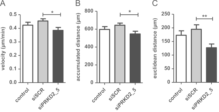
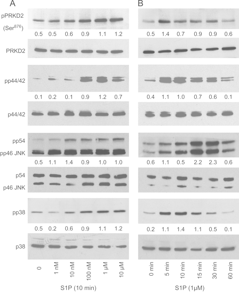
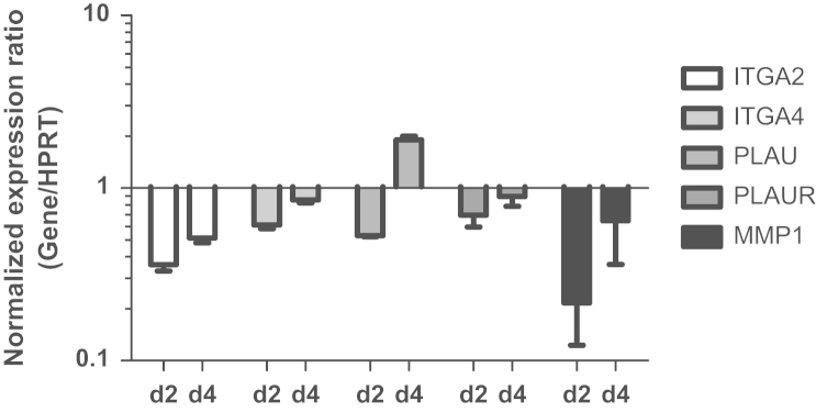
Glioblastoma multiforme (GBM) is the most common malignant brain tumor, which, despite combined modality treatment, reoccurs and is invariably fatal for affected patients. Recently, a member of the serine/threonine protein kinase D (PRKD) family, PRKD2, was shown to be a potent mediator of glioblastoma growth. Here we studied the role of PRKD2 in U87MG glioblastoma cell migration and invasion in response to sphingosine-1-phosphate (S1P), an activator of PRKD2 and a GBM mitogen. Time-lapse microscopy demonstrated that random cell migration was significantly diminished in response to PRKD2 silencing. The pharmacological PRKD family inhibitor CRT0066101 decreased chemotactic migration and invasion across uncoated or matrigel-coated Transwell inserts. Silencing of PRKD2 attenuated migration and invasion of U87MG cells even more effectively. In terms of downstream signaling, CRT0066101 prevented PRKD2 autophosphorylation and inhibited p44/42 MAPK and to a smaller extent p54/46 JNK and p38 MAPK activation. PRKD2 silencing impaired activation of p44/42 MAPK and p54/46 JNK, downregulated nuclear c-Jun protein levels and decreased c-Jun(S73) phosphorylation without affecting the NFκB pathway. Finally, qPCR array analyses revealed that silencing of PRKD2 downregulates mRNA levels of integrin alpha-2 and -4 (ITGA2 and -4), plasminogen activator urokinase (PLAU), plasminogen activator urokinase receptor (PLAUR), and matrix metallopeptidase 1 (MMP1). Findings of the present study identify PRKD2 as a potential target to interfere with glioblastoma cell migration and invasion, two major determinants contributing to recurrence of glioblastoma after multimodality treatment.

Keywords: C-Jun; Glioblastoma; Invasion; MAPK; PRKD2; Sphingosine-1-phosphate.


Figures

Fig. 1
Fig. 1
Efficacy of PRKD2 silencing. Knockdown of PRKD2 expression in U87MG cells was performed by RNA interference using two different siRNA constructs (Oligofectamine was used to transfect 20 nM siRNA). Silencing efficacy was analyzed by (A) qPCR two and four days post transfection and (B) Western blotting three and five days post transfection. Results in (A) represent mean±SD of three different experiments. Results in (B) show one representative Western blot out of five independent experiments. Numbers represent relative optical densities of PRKD2 protein normalized to loading controls.
Fig. 2
Fig. 2
Interference with PRKD2 expression impairs random migration of U87MG cells. For time-lapse microscopy untreated cells and cells transfected with siSCR or siPRKD2_5 were seeded on 24-well tissues. Two days post transfection images were acquired every 20 min for 24 h at five different positions of each well. Cell motility of 50 cells per treatment group was assessed using the Manual Tracking Macro and the Chemotaxis plugin of ImageJ. The (A) mean velocity, (B) mean accumulated distance and (C) the mean Euclidian distance was calculated. *p<0.05, **p<0.01 in comparison to siSCR (1way ANOVA).
Fig. 3
Fig. 3
Interference with PRKD2 expression reduces chemotactic migration and the invasive potential of U87MG cells. Untreated cells, cells transfected with siSCR and siPRKD2_5 and cells treated with 1 µM CRT0066101 were allowed to migrate across uncoated or matrigel-coated Transwell inserts. The lower chamber was loaded with medium containing FCS (10 or 0.5%; v/v) or S1P (1 µM or 2.5 µM; serum free). Cells that migrated to the bottom side of the membrane were fixed with methanol, stained with toluidine blue for 2 min and the membrane was washed twice with water. Cells in six fields from each membrane were counted using an optical microscope (20x magnifications). (A) Representative images of migrated cells. Quantitative analysis of cell migration across (B) uncoated or (C) matrigel-coated Transwell inserts. Data are presented as mean±SEM. Assays were performed in quadruplicate for each condition. ***p<0.001, **p<0.01 in comparison to control or siSCR (1way ANOVA).
Fig. 4
Fig. 4
S1P activates mitogenic signaling cascades in U87MG cells. Cells were grown in 6 well plates and activated with BSA-complexed S1P in a concentration (A; 1 nM–10 µM; 10 min) or time-dependent (B; 1 µM; 5–60 min) manner. Whole cell lysates were separated by SDS-PAGE (10 or 30 µg protein/lane) and transferred to PVDF membrane. Protein levels of phospho-PRKD2 (Ser876), PRKD2, phospho-p44/42 MAPK (pp44/42), p44/42 MAPK, phospho-p38 MAPK (pp38), p38, phospho-p54/46 JNK (p54/46 JNK), and JNK were determined using specific antibodies. Results of one experiment out of three (that provided similar results) are shown.
Fig. 5
Fig. 5
Pharmacological inhibition and silencing of PRKD2 impairs MAPK signaling and c-Jun regulation but does not affect the NFκB pathway. Cells were incubated with (A) CRT0066101 at the indicated concentrations overnight or (B) transfected with scrambled siRNA (S) and siPRKD2_5 (P5) for two days and activated with 1 µM BSA-complexed S1P for the indicated times. Protein levels of PRKD2 and MAPK family member activation were analyzed as described in Fig. 4. Results of one experiment out of three (that provided similar results) are shown. (C) c-Jun expression and activation was analyzed in nontransfected and transfected (siSCR, siPRKD2_5) cells in the absence or presence of S1P (1 µM, 5 min). Nuclear lysates were separated by SDS-PAGE (10 µg protein/lane) and transferred to PVDF membrane. Total c-Jun expression and c-Jun protein activated by phosphorylation at Ser63 or Ser73 were analyzed using specific antibodies. Results of one experiment out of three are shown. (D) Cytosolic and nuclear PRKD2, pPRKD2, and NFκB (p50 and p65) expression was analyzed in nontransfected and transfected (siSCR, siPRKD2_5) cells under unstimulated and stimulated (1 µM S1P for 5 min) conditions. Cytosolic and nuclear protein fractions were separated by SDS-PAGE (10 µg protein/lane) and transferred to PVDF membrane. Immunoreactive bands were detected using rabbit antibodies. One experiment out of two is shown.
Fig. 6
Fig. 6
Silencing of PRKD2 impacts on U87MG gene expression. PRKD2 expression was silenced with siPRKD2_5. Two and four days post transfection target gene expression was analyzed by qPCR using validated primer pairs. Hypoxanthine phosphoribosyltransferase 1 (HPRT) was used as housekeeping gene. Relative gene expression of target genes is presented in relation to mock transfection. Results represent mean±SD from three independent experiments. Gene expression ratios were calculated by REST as described in Materials and methods.

References

    1. Stupp R., Mason W.P., van den Bent M.J., Weller M., Fisher B., Taphoorn M.J., Belanger K., Brandes A.A., Marosi C., Bogdahn U., Curschmann J., Janzer R.C., Ludwin S.K., Gorlia T., Allgeier A., Lacombe D., Cairncross J.G., Eisenhauer E., Mirimanoff R.O. Radiotherapy plus concomitant and adjuvant temozolomide for glioblastoma. N. Engl. J. Med. 2005;352:987–996. - PubMed
    1. Parsons D.W., Jones S., Zhang X., Lin J.C., Leary R.J., Angenendt P., Mankoo P., Carter H., Siu I.M., Gallia G.L., Olivi A., McLendon R., Rasheed B.A., Keir S., Nikolskaya T., Nikolsky Y., Busam D.A., Tekleab H., Diaz L.A., Jr., Hartigan J., Smith D.R., Strausberg R.L., Marie S.K., Shinjo S.M., Yan H., Riggins G.J., Bigner D.D., Karchin R., Papadopoulos N., Parmigiani G., Vogelstein B., Velculescu V.E., Kinzler K.W. An integrated genomic analysis of human glioblastoma multiforme. Science. 2008;321:1807–1812. - PMC - PubMed
    1. The Cancer Genome Atlas Research Network, Comprehensive genomic characterization defines human glioblastoma genes and core pathways, Nature 455 (2008) 1061–1068. - PMC - PubMed
    1. Hadjipanayis C.G., Van Meir E.G. Tumor initiating cells in malignant gliomas: biology and implications for therapy. J. Mol. Med. (Berl) 2009;87:363–374. - PMC - PubMed
    1. Friedl P., Locker J., Sahai E., Segall J.E. Classifying collective cancer cell invasion. Nat. Cell Biol. 2012;14:777–783. - PubMed

Publication types

MeSH terms