. 2013 Jun:111:71-8.
doi: 10.1016/j.exer.2013.03.017. Epub 2013 Apr 3.

Retinal deimination and PAD2 levels in retinas from donors with age-related macular degeneration (AMD)

Affiliations

Retinal deimination and PAD2 levels in retinas from donors with age-related macular degeneration (AMD)

Vera L Bonilha et al. Exp Eye Res. 2013 Jun.

Abstract

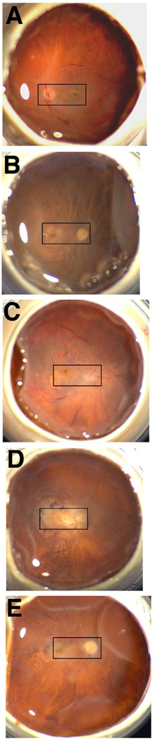
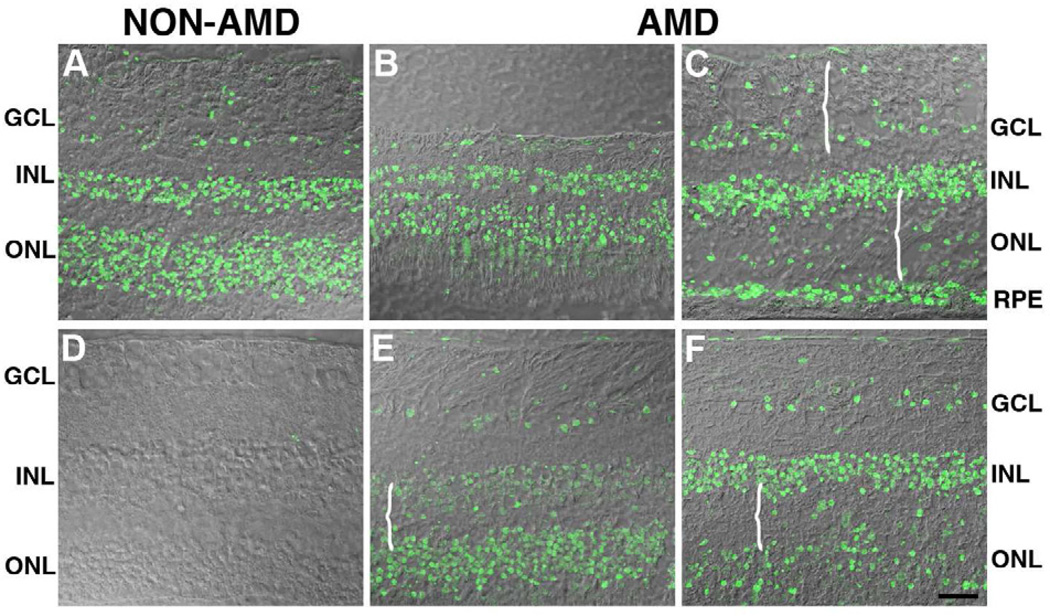
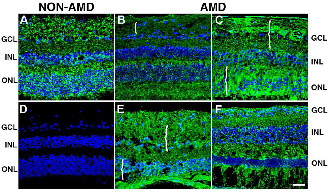
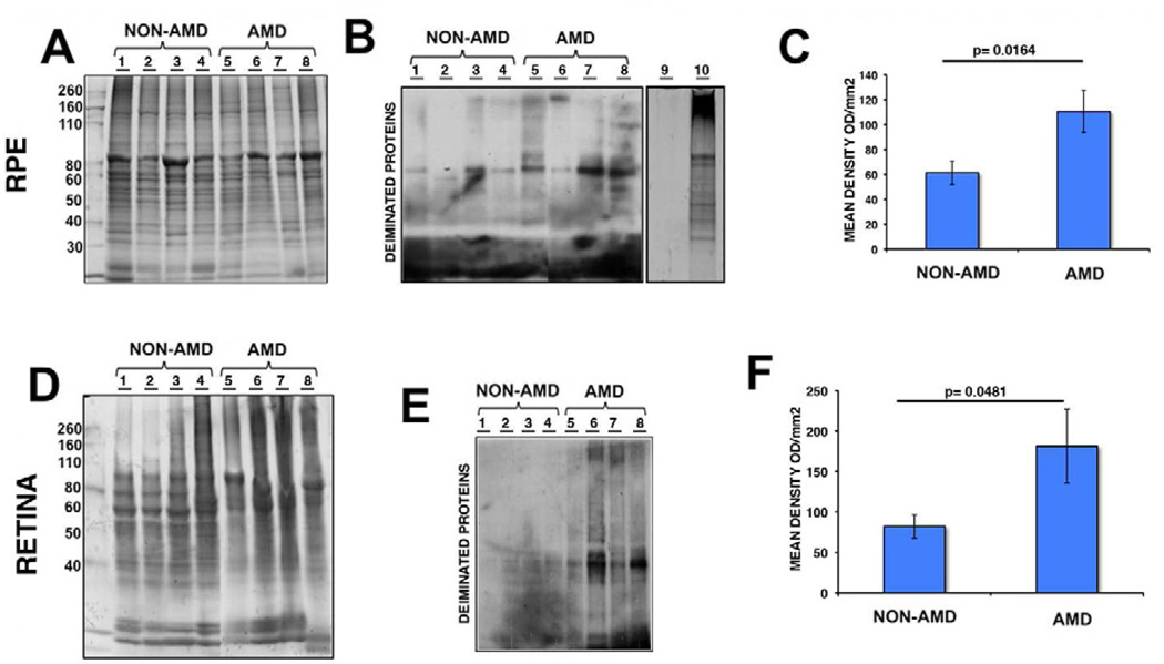
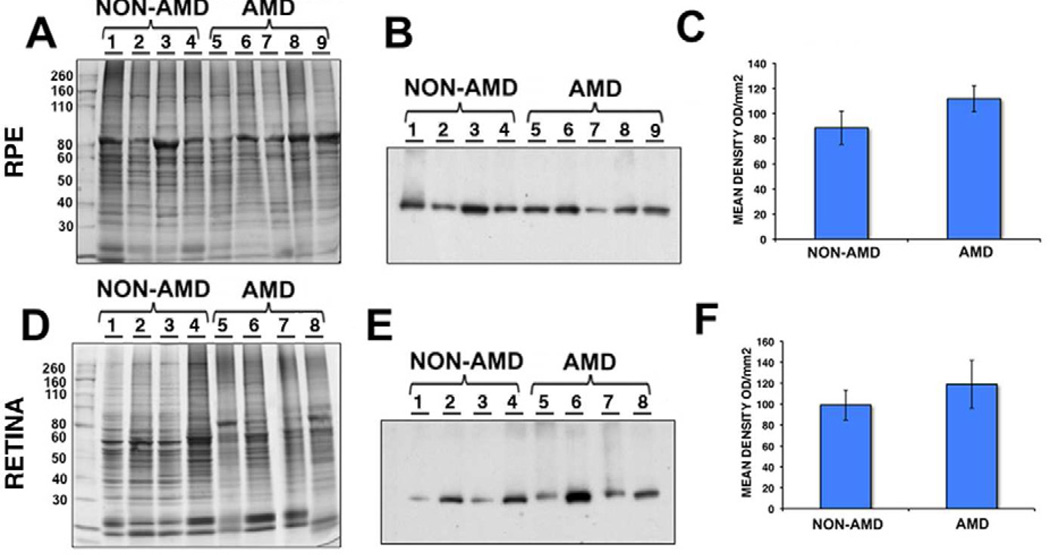
Deimination is a form of protein posttranslational modification carried out by the peptidyl arginine deiminases (PADs) enzymes. PAD2 is the principal deiminase expressed in the retina. Elevated levels of PAD2 and protein deimination are present in a number of human neurological diseases, with or without ocular manifestation. To define the association of deimination with the pathogenesis of age-related macular degeneration (AMD), we studied protein deimination and PAD2 levels in retinas of AMD donor eyes compared to age-matched non-AMD retinas. Eyes from non-AMD and AMD donors were fixed in 4% paraformaldehyde and 0.5% glutaraldehyde in phosphate buffer. Retina and retinal pigment epithelium (RPE) from donor eyes were processed for immunohistochemical detection and western blotting using antibodies to PAD2 and citrulline residues. The ganglion cell, inner plexiform, inner nuclear and outer nuclear layers were labeled by both PAD2 and citrulline antibodies. Changes in the localization of deiminated residues and PAD2 were evident as the retinal layers were remodeled coincident with photoreceptor degeneration in AMD retinas. Immunodetection of either PAD2 or citrulline residues could not be evaluated in the RPE layer due to the high autofluorescence levels in this layer. Interestingly, higher deimination immunoreactivity was detected in AMD retinal lysates. However, no significant changes in PAD2 were detected in the AMD and non-AMD retinas and RPE lysates. Our observations show increased levels of protein deimination but not PAD2 in AMD retinas and RPE, suggesting a reduced rate of turnover of deiminated proteins in these AMD retinas.


Figures

Fig. 1
Fig. 1. In situ imaging of whole AMD and age-matched non-AMD donor eyes
The optic nerve head, macula and retinal veins were visible in the macroscopic fundus image of the non-AMD eye (A). AMD samples displayed typical geographic atrophy (B, D) and exudate accumulation around the macula (C–E). Rectangles highlight the areas selected for histological analysis.
Fig. 2
Fig. 2. Protein deimination localization is similar in the retinas of AMD and non-AMD donors
The levels of protein deimination staining were analyzed in the perimacular area of non-AMD (A) and AMD retinas (B, C, E, F). Immunoreactivity was overlaid on differential contrast images (DIC) of the retina. Analysis of the AMD retinal sections showed that the levels of deiminated proteins observed were similar to the levels observed in non-AMD retinas. Specifically, non-AMD retinas displayed protein deimination in the ganglion cell layer (GCL), inner nuclear layer (INL), outer nuclear layer (ONL). Interestingly, deimination was frequently localized to the nuclei of cells in these layers of both AMD and non-AMD. A disorganized distribution of deiminated proteins was visible in the degenerated areas of the retinas of AMD donors (braces). Non-AMD (D) and AMD (data not shown) retinas labeled only with the secondary antibody did not display any deiminated protein labeling Bar = 40µm.
Fig. 3
Fig. 3. PAD2 levels are similar in the retinas of AMD and non-AMD donors
The levels of PAD2 were analyzed in the perimacular area of non-AMD (A, D) and AMD retinas (B, C, E, F). Nuclei were labeled with TO-PRO3 and are shown in blue to serve as a reference for the retinal layers. Analysis of the AMD retinal sections showed that the levels of PAD2 were similar to the levels observed in non-AMD retinas. Immunolocalization of PAD2 was present in all retinal layers, namely, ganglion cell layer (GCL), inner nuclear layer (INL), outer nuclei layer (ONL). Interestingly, PAD2 was frequently localized to the nuclei of cells in the GCL and INL. A disorganized distribution of PAD2 was visible in the degenerated areas of the retinas of AMD donors (braces) Non-AMD (D) and AMD (data not shown) retinas labeled only with the secondary antibody did not display any deiminated protein labeling. Bar = 40µm.
Fig. 4
Fig. 4. Increased expression of deiminated proteins in RPE and retina lysates of AMD and non-AMD donors
RPE (A–C) and retinas (D–F) from several AMD donors were harvested and lysed. A representative gel is shown stained with Gelcode blue after partial transfer to PVDF membranes to serve as a reference for the load homogeneity of the samples (A, D). The same samples are also shown after immunoreactivity of samples with the anti-citrulline antibody (B, E). Drosophila whole extract (which lacks PADs) before (B, lane 9) and after (B, lane 10) incubation with PAD2 provided control for anti-citrulline antibody immunoreactivity. In C, and F, the mean signal intensity was plotted for AMD and non-AMD samples ± error bars. Protein deimination immunoreactivity was approximately 1.8 fold higher in AMD RPE compared with non-AMD RPE (p = 0. 0164 by Student’s t-test, n= 10). In addition, protein deimination immunoreactivity was approximately 2.2 fold higher in AMD retinas compared with non-AMD retinas (p = 0.0481 by Student’s t-test, n= 7).
Fig. 5
Fig. 5. Similar expression of PAD2 in RPE and retina lysates of AMD and non-AMD donors
RPE (A–C) and retinas (D–F) from AMD donors were harvested and lysed. A representative gel is shown stained with Gelcode blue after partial transfer to PVDF membranes to serve as a reference for the load homogeneity of the samples (A, D). The same samples are also shown after immunoreactivity of samples with the anti-PAD2 antibody (B, E). In C, and F, the mean signal intensity was plotted for AMD and non-AMD samples ± error bars. PAD2 immunoreactivity was approximately 1.2 fold higher in AMD RPE compared with non-AMD RPE (p = 0.2193 by Student’s t-test, n= 10). PAD2 immunoreactivity was also approximately 1.2 fold higher in AMD retinas compared with non-AMD retinas (p = 0.4535 by Student’s t-test, n= 9).

References

    1. Acharya NK, Nagele EP, Han M, Coretti NJ, DeMarshall C, Kosciuk MC, Boulos PA, Nagele RG. Neuronal PAD4 expression and protein citrullination: possible role in production of autoantibodies associated with neurodegenerative disease. J. Autoimmun. 2012;38:369–380. - PubMed
    1. Asaga H, Ishigami A. Protein deimination in the rat brain after kainate administration: citrulline-containing proteins as a novel marker of neurodegeneration. Neurosci. Lett. 2001;299:5–8. - PubMed
    1. Asaga H, Akiyama K, Ohsawa T, Ishigami A. Increased and type II-specific expression of peptidylarginine deiminase in activated microglia but not hyperplastic astrocytes following kainic acid-evoked neurodegeneration in the rat brain. Neurosci. Lett. 2002;326:129–132. - PubMed
    1. Beatty S, Koh H, Phil M, Henson D, Boulton M. The role of oxidative stress in the pathogenesis of age-related macular degeneration. Surv Ophthalmol. 2000;45:115–134. - PubMed
    1. Bando H, Shadrach KG, Rayborn ME, Crabb JW, Hollyfield JG. Clathrin and adaptin accumulation in drusen, Bruch's membrane and choroid in AMD and non-AMD donor eyes. Exp. Eye Res. 2007;84:135–142. - PubMed

Publication types

MeSH terms