Review
. 2013 Sep:36:24-51.
doi: 10.1016/j.preteyeres.2013.03.002. Epub 2013 Apr 3.

Protein sorting, targeting and trafficking in photoreceptor cells

Affiliations
Review

Protein sorting, targeting and trafficking in photoreceptor cells

Jillian N Pearring et al. Prog Retin Eye Res. 2013 Sep.

Abstract

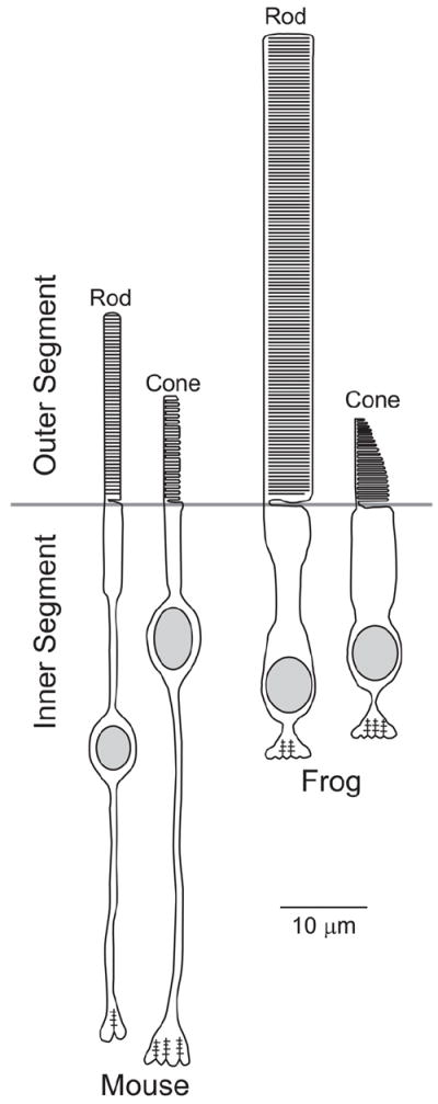
Vision is the most fundamental of our senses initiated when photons are absorbed by the rod and cone photoreceptor neurons of the retina. At the distal end of each photoreceptor resides a light-sensing organelle, called the outer segment, which is a modified primary cilium highly enriched with proteins involved in visual signal transduction. At the proximal end, each photoreceptor has a synaptic terminal, which connects this cell to the downstream neurons for further processing of the visual information. Understanding the mechanisms involved in creating and maintaining functional compartmentalization of photoreceptor cells remains among the most fascinating topics in ocular cell biology. This review will discuss how photoreceptor compartmentalization is supported by protein sorting, targeting and trafficking, with an emphasis on the best-studied cases of outer segment-resident proteins.

Keywords: Cilium; Outer segment; Photoreceptor; Protein targeting; Protein trafficking.


Figures

Fig. 1
Fig. 1
Schematic structures of mouse and frog photoreceptors drawn roughly to scale.
Fig. 2
Fig. 2
Membrane proteins residing in the rod disc rim and outer segment plasma membrane. Rhodopsin is primarily localized in the disc lamellae, though also present in the plasma membrane (not shown). The cGMP-gated (CNG) channel and the Na/Ca/K exchanger are localized to the plasma membrane. The GARP domain of the CNG channel β1-subunit associates with peripherin/rom-1 oligomeric complex located in the disc rim; this interaction is believed to tether the disc to the plasma membrane. Other peripherin/rom-1 oligomers are thought to interact with soluble GARP2, a splice variant of CNGβ1. The ABCA4 transporter is also localized within the disc rim.
Fig. 3
Fig. 3
(A) Drawing of the entire outer segment and the distal inner segment portion of a mammalian cone. As described in the text, mammalian cone outer segments have only minor base-to-distal tapering. (B) A longitudinal section of the distal cone outer segment region, where one disc is continuous with the plasma membrane and several other discs are enclosed. (C) A longitudinal section of the cone outer segment base, where discs exist as contiguous plasma membrane evaginations. Image adapted from (Anderson et al., 1978).
Fig. 4
Fig. 4
Schematic comparison of a primary cilium (left) and a photoreceptor outer segment (right). Drawings to the left of the primary cilium depict tangential sections through the subciliary compartments: basal body, transition zone, axoneme doublets, and axoneme singlets. Electron micrographs represent tangential sections through various ciliary components of the photoreceptor: the basal body of a mouse rod, reproduced from (Sedmak and Wolfrum, 2011); connecting cilium and axoneme doublets of rat rods, reproduced from (Besharse et al., 1985); and the axonemal transition from doublets to singlets of a zebrafish cone. The axonemal transition in the distal outer segment shows two singlets (1 and 5) and 7 doublets adjacent to the OS discs. Bar is 0.33 μm, reproduced from (Insinna et al., 2008).
Fig. 5
Fig. 5
Schematic representation depicting the steps involved in outer segment formation. Outer segment morphogenesis begins when the mother centriole contacts a ciliary vesicle. Upon attachment, axonemal extension from the centriole causes the ciliary vesicle to invaginate and form the ciliary sheath. Fusion with the plasma membrane externalizes the developing outer segment and transforms the outer sheath into the periciliary membrane. Final stages of outer segment morphogenesis consist of disc formation and outer segment extension. (A-E) Electron micrographs of different stages of outer segment morphogenesis correlated with schematic diagram (Sedmak and Wolfrum, 2011). (A) The mother centriole attached to the ciliary vesicle in the cytoplasm of a differentiating photoreceptor. (B) The ciliary vesicle elongates to form the ciliary sheath. (C) The ciliary sheath fuses with the plasma membrane of the inner segment and the newly assembling outer segment emerges on the cell surface. (D-E) The axoneme extends into the outer segment and the first stacks of disc membranes appear.
Fig. 6
Fig. 6
Schematic representations of the open disc, evagination model (A) and the closed disc, vesicle fusion models of rod disc formation (B). (C-D) Electron micrographs of two mouse rod are included to demonstrate the recent publication of images that support either model. Reprinted with permission from (Patil et al., 2012) and (Chuang et al., 2007).
Fig. 7
Fig. 7
Molecular interactions taking place during rhodopsin transport to the outer segment. The figure is reproduced with permission from (Wang et al., 2012). At the trans-Golgi, GTP-bound Arf4 interacts with rhodopsin and they recruit ASAP1 into the ternary complex. ASAP1 likely initiates membrane deformation through its BAR domain while mediating GTP-hydrolysis of Arf4, which then dissociates from the trans-Golgi. ASAP1 then selectively binds Rab11, which also associates with rhodopsin. ASAP1 and Rab11 recruit Rabin8 and Rab8. On rhodopsin transport carriers (RTCs), ASAP1 serves as a scaffold for the Rab11/Rabin8/Rab8 complex, which controls the activation of Rab8. Activated Rab8 regulates RTCs fusion and the delivery of rhodopsin across the membrane diffusion barrier surrounding the connecting cilium.
Fig. 8
Fig. 8
Comparison of R9AP expression with non-targeted and targeted constructs in frog and mouse rods. Immunofluorescent expression of an outer segment targeted construct (a single pass transmembrane domain from the activin receptor fused to a GFP and rhodopsin’s C-terminal 38 amino acids) is exclusive localized to the outer segment in both frog (A) and mouse (D) rods. In contrast, the outer segment un-targeted construct (the same activin-GFP-rhodopsin backbone, but lacking C-terminal VXPX targeting sequence) is localized throughout the frog (B) and mouse (E) rod plasma membrane. As described in the text, the distribution of untargeted construct prefers frog outer segments compared to mouse outer segments. Scale bar is 5 μm (A-C) and 10 μm (D-F).
Fig. 9
Fig. 9
(A) The role of translocating proteins in visual signal transduction. The visual signal is initiated by the photoexcitation of rhodopsin (R*) which activates the heterotrimeric G protein transducin by catalyzing the GDP/GTP exchange on the α-subunit (Gα) followed by Gα dissociation from the βγ-subunit (Gβ and Gγ). Gα bound to GTP stimulates cGMP phosphodiesterase (PDE) leading to decline of cGMP and the onset of the photoresponse. These reactions are illustrated on the upper membrane disc. The reactions responsible for inactivation of R* are illustrated on the lower disc. They include R* phosphorylation by rhodopsin kinase (RK) followed by arrestin (Arr) binding. The RK activity is regulated by the Ca2+-binding protein recoverin (Rec), which binds to and inhibits RK at high Ca2+. The drawings are modified with permission from (Calvert et al., 2006). (B) Schematic illustration of transducin, arrestin and recoverin distribution in dark- and light-adapted rods. The numbers on the left, color-coded to the corresponding translocating proteins, represent the percentage of the proteins found in the outer segments. The subcellular rod compartments are abbreviated on the right: OS – outer segment; IS – inner segment; N – nucleus; ST – synaptic terminal.
Fig.10
Fig.10
Light-dependency of transducin and arrestin translocation in mouse rods. The rod outer segment contents of arrestin and transducin in living anesthetized mice kept in the dark or subjected to 30-40 minutes of continuous illumination of indicated light intensity. Note that the value for the threshold light intensity triggering transducin translocation is updated from ~10,000 R*/rod/sec estimated in an earlier report (Sokolov et al., 2002) to ~4,000 R*/rod/sec directly measured in the subsequent studies (Lobanova et al., 2007; Lobanova et al., 2010). The data for arrestin are taken from (Strissel et al., 2006).
Fig. 11
Fig. 11
The mechanism of transducin translocation. (A) Transducin dissociation from membranes and translocation require the activation and separation of its functional subunits. (B) In light intensity below transducin translocation threshold produces less activated transducin than PDE. (C) In light intensity above transducin translocation threshold there is more activated transducin than PDE. This excess activated transducin, neither retained on the membrane by PDE nor rapidly deactivated by RGS9, dissociates from the membrane to the cytosol and ultimately diffuse out of the rod outer segment. Reproduced with permission from (Arshavsky and Burns, 2012).

References

    1. Adler AJ, Klucznik KM. Proteins and glycoproteins of the bovine interphotoreceptor matrix: composition and fractionation. Exp Eye Res. 1982;34:423–434. - PubMed
    1. Adler AJ, Severin KM. Proteins of the bovine interphotoreceptor matrix: tissues of origin. Exp Eye Res. 1981;32:755–769. - PubMed
    1. Anant JS, Fung BK. In vivo farnesylation of rat rhodopsin kinase. Biochem Biophys Res Commun. 1992;183:468–473. - PubMed
    1. Anant JS, Ong OC, Xie HY, Clarke S, O’Brien PJ, Fung BK. In vivo differential prenylation of retinal cyclic GMP phosphodiesterase catalytic subunits. J Biol Chem. 1992;267:687–690. - PubMed
    1. Anderson DH, Fisher SK. Disc shedding in rodlike and conelike photoreceptors of tree squirrels. Science. 1975;187:953–955. - PubMed

Publication types