

Spontaneous hepatic rupture in a patient with peliosis hepatis: A report of one case
- PMID: 23562904
- PMCID: PMC3731704
- DOI: 10.1016/j.ijscr.2013.01.030
Spontaneous hepatic rupture in a patient with peliosis hepatis: A report of one case
Abstract
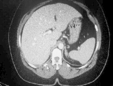
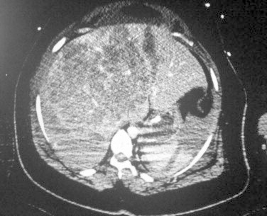
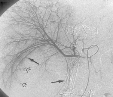
Introduction: Liver rupture is a serious event that is most commonly due to blunt abdominal trauma. We present a case of peliosis hepatis in a patient admitted for acute pyelonephritis who developed hemoperitoneum due to spontaneous hepatic rupture from this rare liver condition.
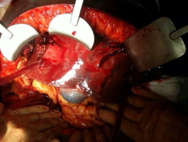
Presentation of case: We report a 44 year-old woman who presented to our hospital with acute pyelonephrititis and hemoperitoneum due to spontaneous hepatic rupture from peliosis hepatis. Physicians should be aware of this rare condition in patients who present with non-traumatic hepatic rupture with hemoperitoneum.
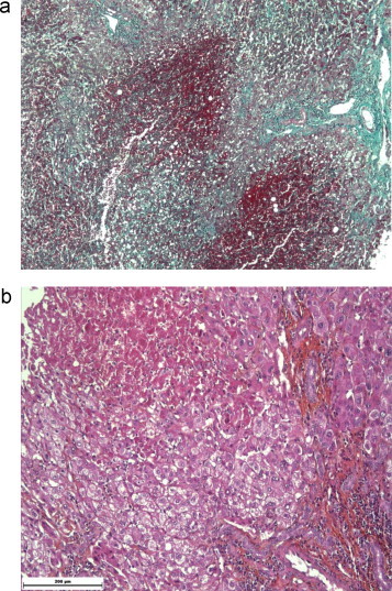
Discussion: PH should be considered in all patients with known risk factors who present with typical morphological changes or a hepatic mass, especially when the cause of sudden intraperitoneal hemorrhage is obscure.
Conclusion: Peliosis hepatis is most often asymptomatic and an incidental finding at autopsy. In symptomatic patients, surgery should be reserved for those patients whose hemorrhage is-life-threatening. Familiarity with the imaging characteristics can help in earlier diagnosis of peliosis hepatis.
Copyright © 2013 Surgical Associates Ltd. Published by Elsevier Ltd. All rights reserved.
Figures
References
-
- Nelson E.W., Archibald L., Albo D. Spontaneous hepatic rupture in pregnancy. American Journal of Surgery. 1977;134:817–820. - PubMed
-
- Adam G., Lesser T., Neumann R. Liver rupture in peliosis hepatis. Zentralblatt fur Chirurgie. 1991;116:399–403. - PubMed
-
- Smith L.G., Moise K.J., Dildy G.A., Carpentier R.J. Spontaneous rupture of liver during pregnancy: current teraphy. Obstetrics and Gynecology. 1991;77:171–175. - PubMed
-
- Hayward S.R., Lucas C.E., Ledgerwood A.M. Recurrent spontaneous intrahepatic hemorrhage from peliosis hepatis. Archives of Surgery. 1991;126:782–783. - PubMed
LinkOut - more resources
Full Text Sources
Other Literature Sources
Research Materials
