

Extraosseous Ewing sarcoma of the vagina: a rare entity
- PMID: 23563009
- PMCID: PMC6078614
- DOI: 10.5144/0256-4947.2013.182
Extraosseous Ewing sarcoma of the vagina: a rare entity
Abstract
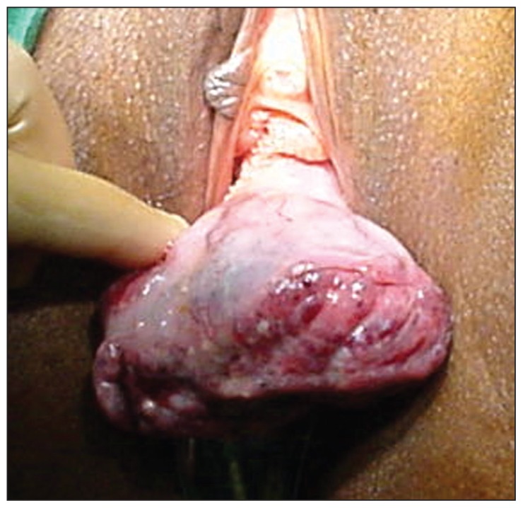
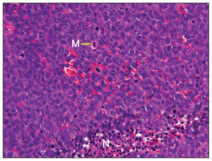
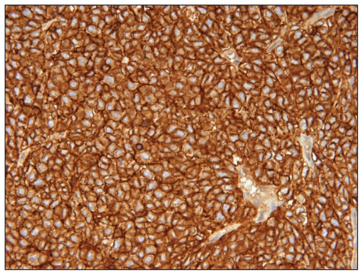
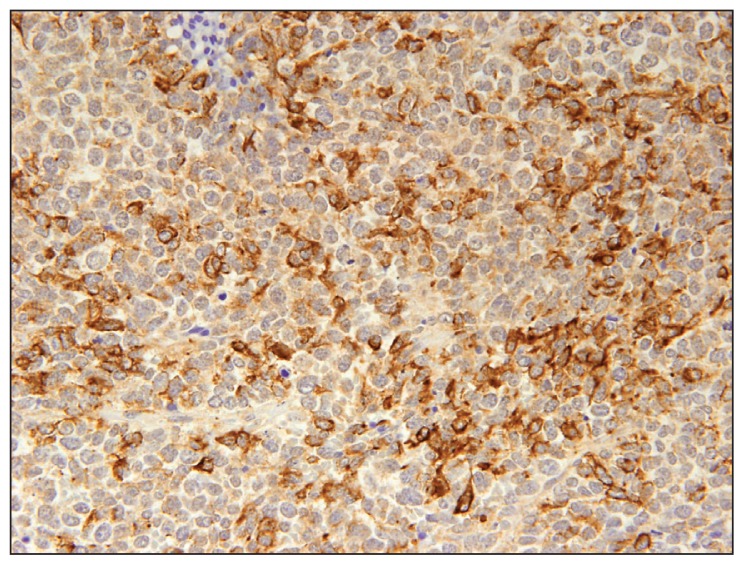
Ewing sarcoma, a highly malignant neoplasm of the bone, usually occurs during childhood. About 15% are extraosseous. The Ewing family of tumors (EFTs) are extremely rare in the vagina. A 40-year literature review from 1970 to 2010 revealed only nine cases. A 32-year-old woman presented with a painless vaginal mass. A wide excision was performed. Histopathology, immunohistochemistry and molecular studies confirmed extraosseous vaginal Ewing sarcoma. Despite aggressive chemotherapy with a good initial response, she developed local recurrence and metastasis to the spine and pelvis and succumbed 22 months later. A previous infiltrating ductal breast cancer, treated and in remission complicated the picture. We present the tenth case of vaginal Ewing sarcoma and the fourth to be confirmed by molecular studies. We stress the importance of molecular techniques in definitely diagnosing EFTs, especially those arising at unusual sites, particularly in the context of a previous diagnosis of breast cancer.
Figures

Similar articles
-
Primary vaginal Ewing sarcoma with uterine fibroid: A case report.Medicine (Baltimore). 2020 Jul 2;99(27):e20859. doi: 10.1097/MD.0000000000020859. Medicine (Baltimore). 2020. PMID: 32629673 Free PMC article.
-
Extraosseous Ewing sarcoma of the vagina.Obstet Gynecol. 2000 Nov;96(5 Pt 2):832-4. doi: 10.1016/s0029-7844(00)01033-4. Obstet Gynecol. 2000. PMID: 11094227
-
Primary vaginal extraosseous Ewing sarcoma/primitive neuroectodermal tumor with cranial metastasis.J Chin Med Assoc. 2009 Jun;72(6):332-5. doi: 10.1016/S1726-4901(09)70381-8. J Chin Med Assoc. 2009. PMID: 19541570
-
Primary Ewing Sarcoma Presenting as a Vulvar Mass in an Adolescent: Case Report and Review of Literature.J Pediatr Adolesc Gynecol. 2015 Dec;28(6):e179-83. doi: 10.1016/j.jpag.2015.04.004. Epub 2015 Apr 14. J Pediatr Adolesc Gynecol. 2015. PMID: 26211932 Review.
-
Ewing sarcoma in an infant and review of the literature.Turk J Pediatr. 2019;61(5):760-764. doi: 10.24953/turkjped.2019.05.016. Turk J Pediatr. 2019. PMID: 32105009 Review.
Cited by
-
Peripheral primitive neuroendocrine tumor of the chest wall-A case report with pathological correlation.Radiol Case Rep. 2018 Feb 5;13(2):392-396. doi: 10.1016/j.radcr.2018.01.003. eCollection 2018 Apr. Radiol Case Rep. 2018. PMID: 29904480 Free PMC article.
-
Importance of studying primitive neuroectodermal tumors and extraosseous Ewings sarcoma of the vagina and vulva.Oncol Lett. 2021 Feb;21(2):171. doi: 10.3892/ol.2021.12432. Epub 2021 Jan 4. Oncol Lett. 2021. PMID: 33552288 Free PMC article. Review.
-
Ewing Sarcoma of the Vagina: A Rare Clinical Entity.Cureus. 2024 Mar 20;16(3):e56550. doi: 10.7759/cureus.56550. eCollection 2024 Mar. Cureus. 2024. PMID: 38646356 Free PMC article.
-
Extraskeletal Ewing sarcoma of thyroid gland: A case report.Zhong Nan Da Xue Xue Bao Yi Xue Ban. 2021 May 28;46(5):558-564. doi: 10.11817/j.issn.1672-7347.2021.200161. Zhong Nan Da Xue Xue Bao Yi Xue Ban. 2021. PMID: 34148894 Free PMC article. Chinese, English.
-
Primary vaginal Ewing sarcoma with uterine fibroid: A case report.Medicine (Baltimore). 2020 Jul 2;99(27):e20859. doi: 10.1097/MD.0000000000020859. Medicine (Baltimore). 2020. PMID: 32629673 Free PMC article.
References
-
- Gaona-Luviano P, Unda-Franco E, González-Jara L, Romero P, Medina-Franco H. Primitive neuroectodermal tumor of the vagina. Gynaecol Oncol. 2003;91:456–8. - PubMed
-
- Petkovic M, Zamolo G, Muhvic D. The first report of extraosseous Ewings sarcoma in the rectovaginal septum. Tumori. 2002;88:345–6. - PubMed
-
- Vang R, Taubenberger JK, Mannion CM. Primary vulvar and vaginal extra-osseous Ewings sarcoma/ peripheral neuroectodermal tumor: diagnostic confirmation with CD99 immunostaining and reverse transcriptase-polymerase chain reaction. Int J Gynecol Pathol. 2000;19:103–9. - PubMed
-
- Farley J, O’Boyle JD, Heaton J, Remmenga S. Extraosseous Ewings sarcoma of the vagina. Obstet Gynecol. 2000;96:832–4. - PubMed
Publication types
MeSH terms
LinkOut - more resources
Full Text Sources
Other Literature Sources
Medical