. 2013 May 1;41(10):5210-22.
doi: 10.1093/nar/gkt223. Epub 2013 Apr 5.

Ribosomal protein S7 regulates arsenite-induced GADD45α expression by attenuating MDM2-mediated GADD45α ubiquitination and degradation

Affiliations

Ribosomal protein S7 regulates arsenite-induced GADD45α expression by attenuating MDM2-mediated GADD45α ubiquitination and degradation

Ming Gao et al. Nucleic Acids Res. .

Abstract

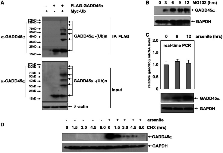
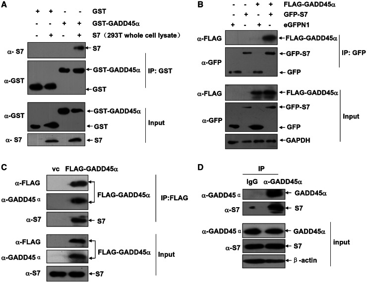
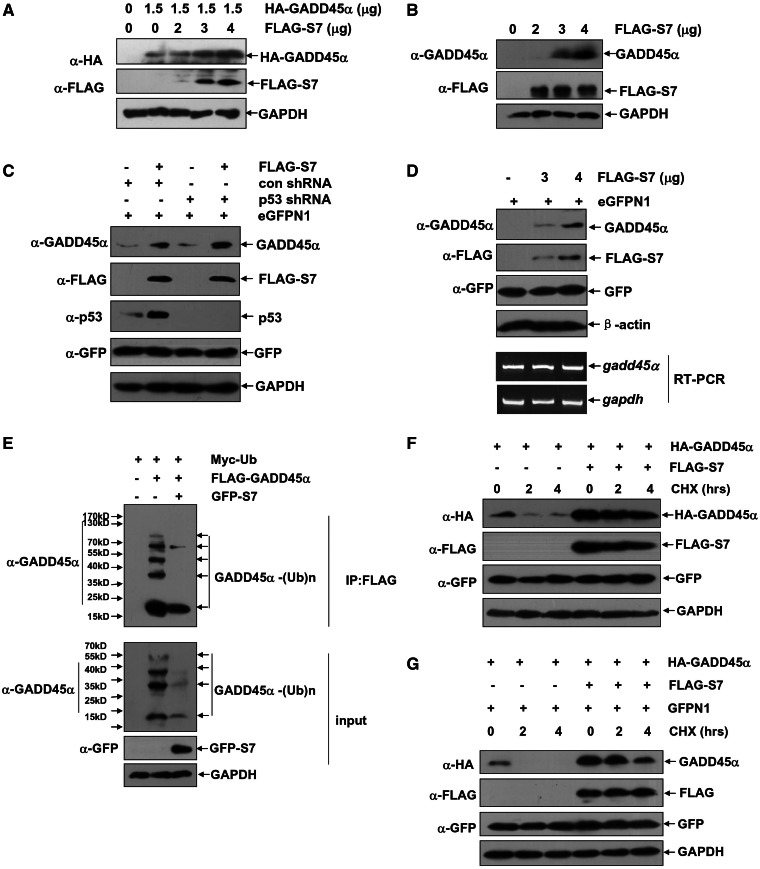
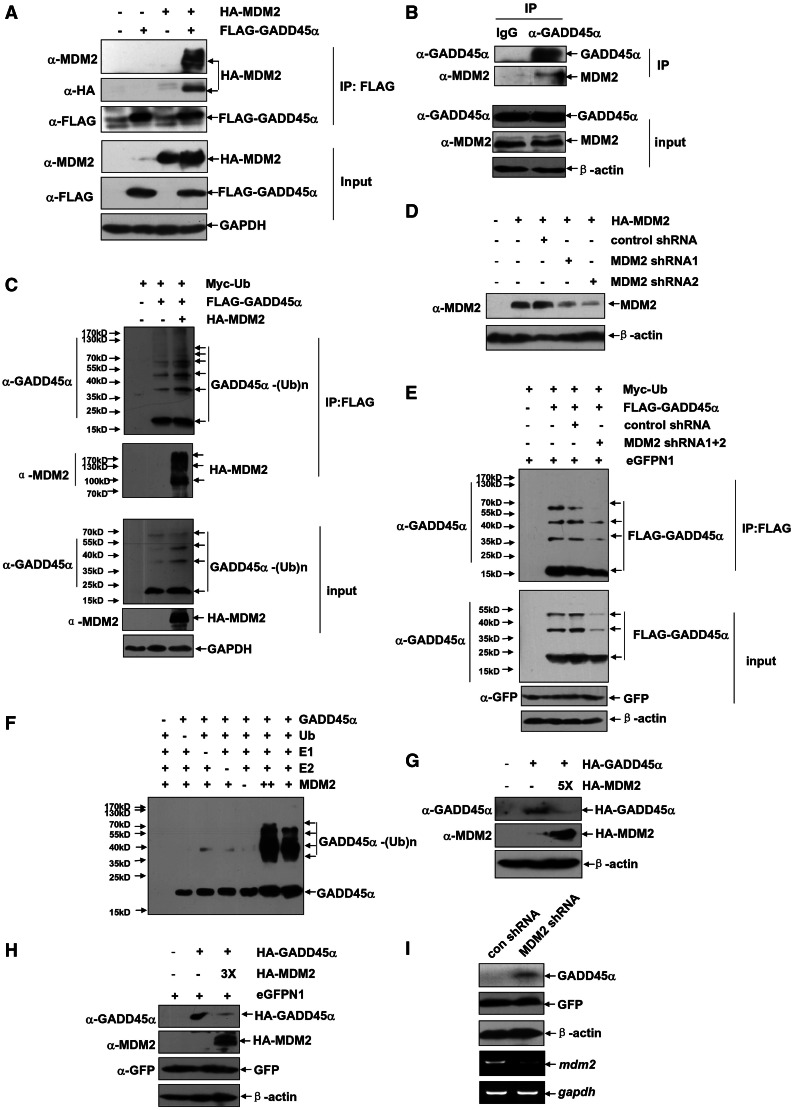
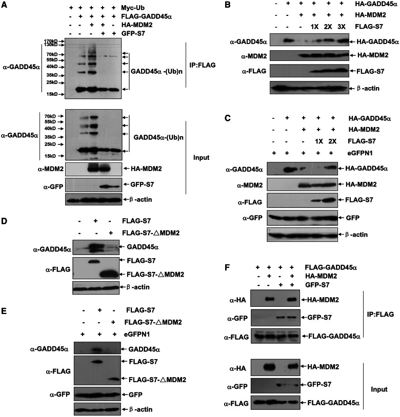
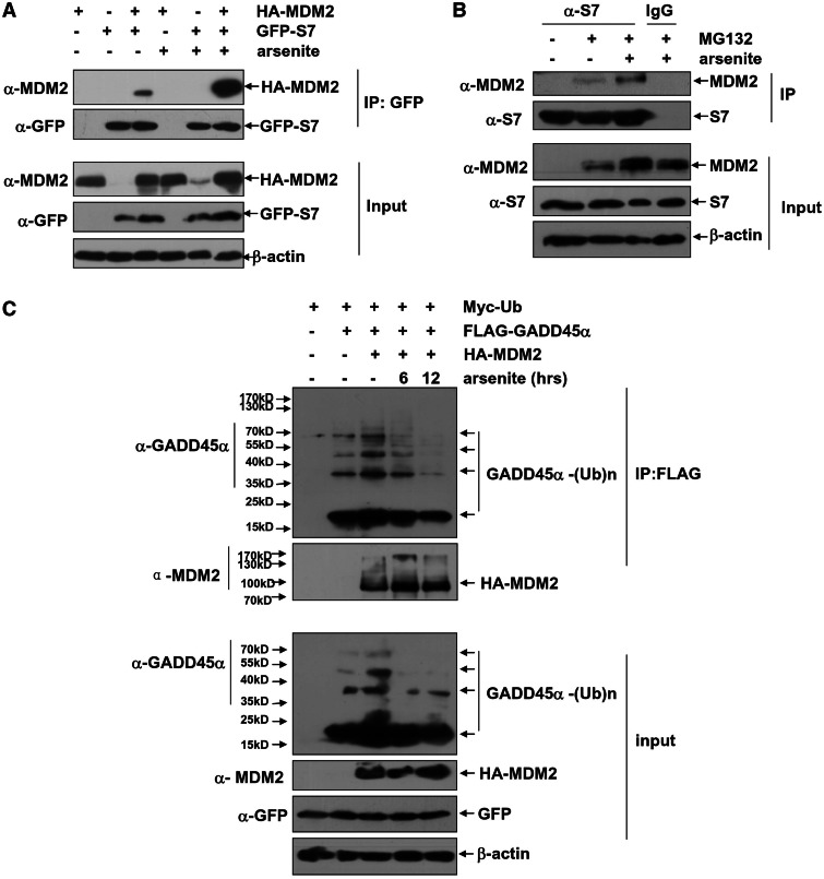
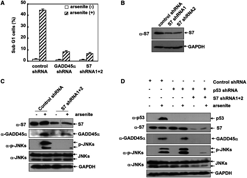
The stress-responding protein, GADD45α, plays important roles in cell cycle checkpoint, DNA repair and apoptosis. In our recent study, we demonstrate that GADD45α undergoes a dynamic ubiquitination and degradation in vivo, which process can be blocked by the cytotoxic reagent, arsenite, resulting in GADD45α accumulation to activate JNKs cell death pathway, thereby revealing a novel mechanism for the cellular GADD45α functional regulation. But the factors involved in GADD45α stability modulations are unidentified. Here, we demonstrated that MDM2 was an E3 ubiquitin ligase for GADD45α. One of MDM2-binding partner, ribosomal protein S7, interacted with and stabilized GADD45α through preventing the ubiquitination and degradation of GADD45α mediated by MDM2. This novel function of S7 is unrelated to p53 but seems to depend on S7/MDM2 interaction, for the S7 mutant lacking MDM2-binding ability lost its function to stabilize GADD45α. Further investigations indicated that arsenite treatment enhanced S7-MDM2 interaction, resulting in attenuation of MDM2-dependent GADD45α ubiquitination and degradation, thereby leading to GADD45α-dependent cell death pathway activation. Silencing S7 expression suppressed GADD45α-dependent cytotoxicity induced by arsenite. Our findings thus identify a novel function of S7 in control of GADD45α stabilization under both basal and stress conditions and its significance in mediating arsenite-induced cellular stress.


Figures

Figure 1.
Figure 1.
GADD45α is a liable protein, and its proteasome-dependent degradation is blocked by arsenite in the HepG2 cells. (A) HepG2 cells were transfected with combination of the expression plasmids encoding FLAG–GADD45α or Myc–Ub as indicated. Cell extracts were immunoprecipitated with anti-FLAG antibody, and the ubiquitination of GADD45α was detected with anti-GADD45α antibody. (B) HepG2 cells were treated with MG132 (10 µM) for the indicated time, and then the levels of GADD45α were detected. (C) HepG2 cells were treated with arsenite (20 µM) for the indicated time, and then the induction of gadd45α mRNA and protein levels was detected by real-time PCR or western-blot assays, respectively. (D) HepG2 cells were left untreated or treated with arsenite (20 µM) for 12 h and then subjected to CHX (20 µM) exposure at the indicated time after arsenite withdrawal. The degradation of GADD45α was detected by western-blot assay.
Figure 2.
Figure 2.
S7 interacts with GADD45α. (A) The purified GST–GADD45α or GST proteins and 293T whole-cell lysate with high levels of S7 expression were incubated together, and then GST-pull-down assay was performed to examine the in vitro interaction between GADD45α and S7. (B) 293T cells were either transfected with the expression plasmid encoding FLAG–GADD45α or in combination with GFP–S7 construct or eFGPN1 vector. Cell lysate was immunoprecipitated with anti-GFP antibody, and then the immunoprecipitants were probed with anti-GFP and anti-FLAG antibodies. (C) HepG2 cells were transfected with FLAG–GADD45α construct or its control vector. Cell lysate was immunoprecipitated with anti-FLAG antibody, and the immunoprecipitants were probed with anti-FLAG, anti-GADD45α and anti-S7 antibodies. (D) HepG2 cells were treated with arsenite (20 µM) for 12 h, and then cell lysate was immunoprecipitated with anti-GADD45α antibody or rabbit IgG. The immunoprecipitants were probed with anti-GADD45α and anti-S7 antibodies.
Figure 3.
Figure 3.
S7 upregulates GADD45α protein stability by attenuating GADD45α ubiquitination and degradation under physiological conditions. (A) HepG2 cells were left untreated or transfected with a single dose of expression plasmid encoding HA–GADD45α (1.5 µg) with or without combination of increasing amount of FLAG–S7 construct (2, 3 and 4 µg). The expression levels of the exogenous GADD45α were detected with anti-HA antibody. (B) HepG2 cells were transfected with increasing amount of FLAG–S7 construct (2, 3 and 4 µg). The expression levels of the endogenous GADD45α were detected with anti-GADD45α antibody. (C) HepG2 cells were transfected with p53 shRNA or its control vector with or without combination of FLAG–S7 construct (3 µg). The expression levels of the endogenous GADD45α and p53 were detected. The cells were also co-transfected with eGFPN1 vector (100 ng) and the expression level of GFP served as the control for both transfection efficiency and global translation in HepG2 cells. (D) H1299 cells were transfected with increasing amount of FLAG–S7 construct (3 and 4 µg). The expression levels of the endogenous GADD45α and the transcription of gadd45α mRNA were detected by western-blot and RT–PCR assay, respectively. (E) HepG2 cells were transfected with Myc–Ub and FLAG–GADD45α constructs with or without combination of the expression plasmid encoding GFP–S7. The cell lysate was immunoprecipitated with anti-FLAG antibody, and then the ubiquitination of GADD45α was detected with anti-GADD45α antibody. (F and G) HepG2 (F) or H1299 (G) cells were transfected with the expression plasmid encoding HA–GADD45α (1 µg) with or without combination of FLAG–S7 construct (3 µg). Then the cells were subjected to CHX (20 µM) exposure at the indicated time, and the degradation of GADD45α was detected with anti-HA antibody.
Figure 4.
Figure 4.
MDM2 functions as an E3 ubiquitin ligase for GADD45α. (A) 293T cells were either transfected with the expression plasmid encoding FLAG–GADD45α or in combination with HA–MDM2 construct. The cells were subjected to MG132 (10 µM) treatment for 4 h before harvesting. Then cell lysate was immunoprecipitated with anti-FLAG antibody, and the immunoprecipitants were probed with anti-FLAG, anti-HA and anti-MDM2 antibodies. (B) HepG2 cells were treated with arsenite (20 µM) for 12 h, and then cell lysate was immunoprecipitated with anti-GADD45α antibody or rabbit IgG. The immunoprecipitants were probed with anti-GADD45α and anti-MDM2 antibodies. (C) HepG2 cells were transfected with Myc–Ub and FLAG–GADD45α constructs with or without combination of the expression plasmid encoding HA–MDM2. Cell lysate was immunoprecipitated with anti-FLAG antibody, and then the ubiquitination of GADD45α was detected with anti-GADD45α antibody. (D) HepG2 cells were transfected with the HA–MDM2 construct with or without combination of control shRNA or MDM2 shRNAs. Then the efficiency of MDM2 shRNAs was determined by western-blot assay. (E) HepG2 cells were transfected with Myc–Ub and FLAG–GADD45α constructs with or without combination of control shRNA or MDM2 shRNAs mixture. Cell lysate was immunoprecipitated with anti-FLAG antibody, and then the ubiquitination of GADD45α was detected with anti-GADD45α antibody. (F) The purified GADD45α protein (250 ng) was incubated in the in vitro ubiquitination reaction buffer with E1 (20 ng), E2 mixture (125 ng), MDM2 (100 or 400 ng) and ubiquitin (600 ng) for 40 min at 37°C. Then the ubiquitination of GADD45α was detected by immunoblotting with the anti-GADD45α antibody. (G) HepG2 cells were transfected with HA–GADD45α construct (0.25 µg) with or without combination of overdosed HA–MDM2 expression plasmid (1.25 µg). Then the expression levels of GADD45α were detected. (H) H1299 cells were transfected with HA–GADD45α construct (0.5 µg) with or without combination of overdosed HA–MDM2 expression plasmid (1.5 µg). Then the expression levels of GADD45α were detected. (I) H1299 cells were left untreated or transfected with MDM2 shRNA. Then the expression levels of endogenous GADD45α and mdm2 mRNA were detected.
Figure 5.
Figure 5.
S7 attenuates MDM2-mediated GADD45α ubiquitination and degradation. (A) HepG2 cells were transfected with Myc–Ub expression plasmid in combination of FLAG–GADD45α, HA–MDM2 or GFP–S7 constructs as indicated. Then the ubiquitination of GADD45α was detected with anti-GADD45α antibody. (B) HepG2 cells were left untreated or transfected with HA–GADD45α construct (0.25 µg) with or without combination of HA–MDM2 expression plasmid (1.25 µg) and increasing amount of FLAG–S7 construct (1.25, 2.5 and 3.75 µg, respectively). Then the levels of GADD45α were detected. (C) H1299 cells were left untreated or transfected with HA–GADD45α construct (0.5 µg) with or without combination of HA–MDM2 expression plasmid (1.5 µg) and increasing amount of FLAG–S7 construct (1.5 and 3.0 µg, respectively). Then the levels of GADD45α were detected. (D and E) HepG2 (D) or H1299 (E) cells were transfected with equal amount of FLAG–S7 or FLAG–S7–ΔMDM2 constructs (3 µg), and then the levels of the endogenous GADD45α were detected. (F) 293 T cells were transfected with combination of FLAG–GADD45α, HA–MDM2 or GFP–S7 constructs as indicated. The cells were subjected to MG132 (10 µM) treatment for 4 h before harvesting. Then cell lysate was immunoprecipitated with anti-FLAG antibody, and the immunoprecipitants were probed with anti-FLAG, anti-GFP or anti-MDM2 antibodies.
Figure 6.
Figure 6.
Arsenite exposure enhances S7–MDM2 interaction, which subsequently abrogates GADD45α ubiquitination. (A) HepG2 cells were either transfected with HA–MDM2 or in combination with GFP–S7. Then the cells were left untreated or exposed to arsenite (20 µM) for 8 h. Cell lysate was immunoprecipitated with anti-GFP antibody, and the immunoprecipitants were probed with anti-GFP and anti-MDM2 antibodies. (B) HepG2 cells were left untreated or pre-treated with MG132 (10 µM) for 6 h followed by exposure to arsenite (20 µM) for 8 h. Cell lysate was immunoprecipitated with anti-S7 antibody or the mouse IgG, and then the immunoprecipitants were probed with anti-S7 and anti-MDM2 antibodies. (C) HepG2 cells were transfected with Myc–Ub expression plasmid with or without combination of FLAG–GADD45α or HA–MDM2 constructs. Then the transfected cells were left untreated or exposed to arsenite (20 µM) for the time indicated. Cell lysate was immunoprecipitated with anti-FLAG antibody, and then the ubiquitination of GADD45α was detected with anti-GADD45α antibody.
Figure 7.
Figure 7.
Knockdown S7 levels impair GADD45α-dependent cell apoptotic pathway activation in the arsenite-treated HepG2 cells. (A) HepG2 cells were transfected with the control shRNA, p53 shRNA or combination of S7 shRNA1 and 2. The cell death incidence was detected by flow cytometric assay 24 h after arsenite exposure. And the cells accumulated in the sub G1 phase were presented. (B) HepG2 cells were transfected with either the control shRNA or the individual S7 shRNA1 or S7 shRNA 2. Then the efficiency of each shRNA was determined with anti-S7 antibody. (C) HepG2 cells were transfected with either the control shRNA or S7 shRNA mixture. Then the cells were left untreated or exposed to arsenite (20 µM) for 12 h. The induction of GADD45α expression and JNKs activation was detected. (D) HepG2 cells were transfected with either the control shRNA or p53 shRNA with or without S7 shRNA mixture. Then the cells were treated as described in C. The induction of GADD45α and p53 expression, as well as JNKs activation, was detected.

Similar articles

Cited by

References

    1. Moskalev AA, Smit-McBride Z, Shaposhnikov MV, Plyusnina EN, Zhavoronkov A, Budovsky A, Tacutu R, Fraifeld VE. Gadd45 proteins: Relevance to aging, longevity and age-related pathologies. Ageing Res. Rev. 2012;11:51–66. - PMC - PubMed
    1. Gao M, NIng G, Chuanshu H, Lun S. Diverse roles of GADD45a in stres signaling. Curr. Protein Pep. Sci. 2009;10:388–394. - PubMed
    1. Wang XW, Zhan Q, Coursen JD, Khan MA, Kontny HU, Yu L, Hollander MC, Oonnor PM, Fornace AJ, Harris CC. GADD45 induction of a G2/M cell cycle checkpoint. Proc. Natl Acad. Sci. USA. 1999;96:3706–3711. - PMC - PubMed
    1. Barreto G, Schafer A, Marhold J, Stach D, Swaminathan SK, Handa V, Doderlein G, Maltry N, Wu W, Lyko F, et al. Gadd45a promotes epigenetic gene activation by repair-mediated DNA demethylation. Nature. 2007;445:671–675. - PubMed
    1. Rai K, Huggins IJ, James SR, Karpf AR, Jones DA, Cairns BR. DNA Demethylation in Zebrafish Involves the Coupling of a Deaminase, a Glycosylase, and Gadd45. Cell. 2008;135:1201–1212. - PMC - PubMed

Publication types

MeSH terms