. 2013 Jun;123(6):2380-94.
doi: 10.1172/JCI66904.

Smoothened is a master regulator of adult liver repair

Affiliations

Smoothened is a master regulator of adult liver repair

Gregory A Michelotti et al. J Clin Invest. 2013 Jun.

Abstract

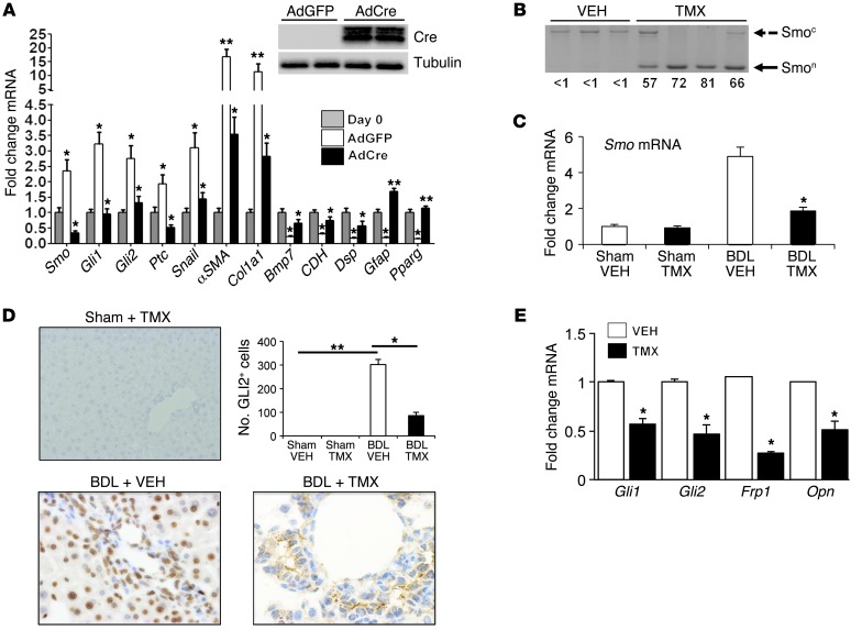
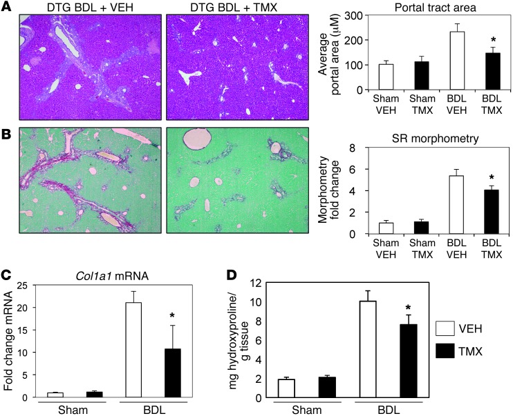
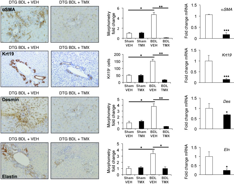
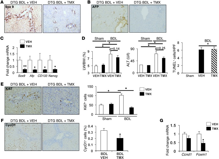
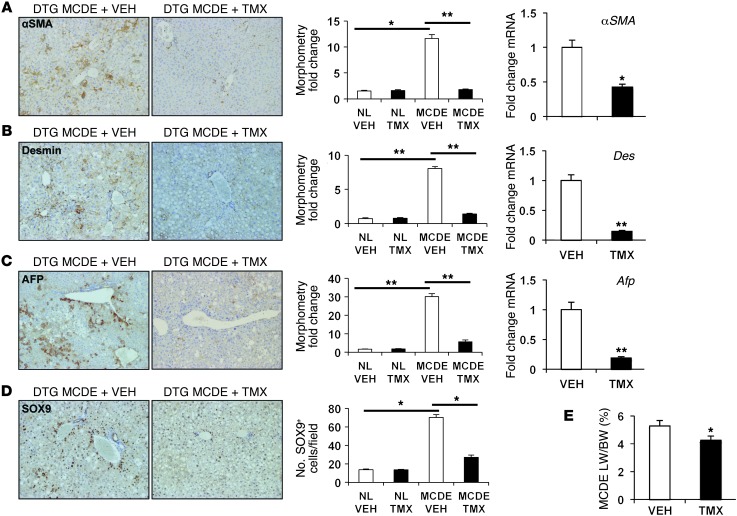
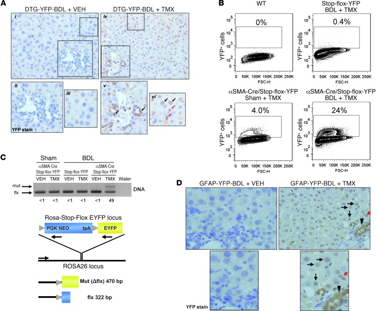
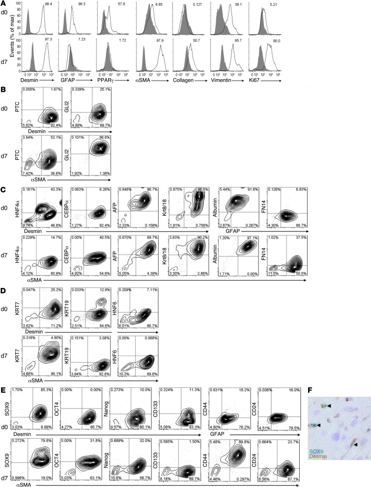
When regenerative processes cannot keep pace with cell death, functional epithelia are replaced by scar. Scarring is characterized by both excessive accumulation of fibrous matrix and persistent outgrowth of cell types that accumulate transiently during successful wound healing, including myofibroblasts (MFs) and progenitors. This suggests that signaling that normally directs these cells to repair injured epithelia is deregulated. To evaluate this possibility, we examined liver repair during different types of liver injury after Smoothened (SMO), an obligate intermediate in the Hedgehog (Hh) signaling pathway, was conditionally deleted in cells expressing the MF-associated gene, αSMA. Surprisingly, blocking canonical Hh signaling in MFs not only inhibited liver fibrosis but also prevented accumulation of liver progenitors. Hh-sensitive, hepatic stellate cells (HSCs) were identified as the source of both MFs and progenitors by lineage-tracing studies in 3 other strains of mice, coupled with analysis of highly pure HSC preparations using flow cytometry, immunofluorescence confocal microscopy, RT-PCR, and in situ hybridization. The results identify SMO as a master regulator of hepatic epithelial regeneration based on its ability to promote mesenchymal-to-epithelial transitions in a subpopulation of HSC-derived MFs with features of multipotent progenitors.


Figures

Figure 1
Figure 1. Conditional disruption of SMO in αSMA+ cells inhibits Hh signaling in MFs.
(A) HSCs were isolated from Smo-flox mice. AdGFP or AdCre (MOI 25) were added on culture day 4, and cells were harvested 3 days later. Effects on gene expression were assessed by qRT-PCR. Results are normalized to day 0 HSCs. *P < 0.05. Inset shows representative Western blot for expression of Cre and tubulin. (B) Oligonucleotides for the floxed SMO allele (refs. , ; Supplemental Table 3) were used to distinguish the unrearranged allele (Smoc; 1.7 Kb) from the Cre-recombined null allele (Smon; 0.5 Kb). Genomic DNA was isolated from livers of DTG mice treated either with vehicle (VEH, n = 3) or TMX (n = 4) from day 4–10 after BDL, and Smon and Smoc amplicons were amplified. Quantification of the amplicons was performed by densitometry to assess the efficiency of Cre-mediated recombination. Results are expressed as Smon/Smon + Smoc x 100, shown below each lane. (C) Total RNA was isolated from livers of vehicle- and TMX-treated DTG mice 14 days after sham surgery or BDL. SMO expression was assessed by qRT-PCR, and results are expressed as fold over sham plus vehicle control. *P < 0.05. (D) Representative immunohistochemistry for hepatic GLI2. GLI2+ cells in vehicle-treated and TMX-treated DTG mice were quantified in sham and BDL groups and represented as number of positive cells per field (n = 11/group; original magnification, ×20 [Sham]; ×63 [BDL]). *P < 0.05, **P < 0.01. (E) Total RNA was isolated from livers of vehicle- and TMX-treated DTG mice 14 days after BDL and analyzed by qRT-PCR for expression of Hh-regulated genes. Results are expressed as fold over vehicle-treated group. *P < 0.05.
Figure 2
Figure 2. Blocking Hh signaling inhibits portal tract expansion and fibrosis in injured livers.
(A) H&E staining of liver sections from representative vehicle- and TMX-treated DTG mice 14 days after sham surgery or BDL (original magnification, ×4). Portal tract size was evaluated by morphometric analysis of H&E-stained sections from all mice (n = 4 mice in sham vehicle; n = 4 mice in sham TMX; n = 11 mice in BDL vehicle; n = 11 mice BDL TMX), expressed as mean ± SEM of portal tract size, and graphed relative to vehicle-treated sham-operated mice. *P < 0.05 vs. vehicle-treated sham group. (B) Other sections were stained with Sirius red/fast green to demonstrate fibrosis (original magnification, ×4). Morphometric data are displayed as mean ± SEM. *P < 0.05 vs. vehicle-treated DTG mice after BDL. (C) Total liver RNA was isolated, and Col1a1 mRNA levels were analyzed by qRT-PCR. Results are expressed as fold over vehicle-treated sham-operated DTG group. *P < 0.05 vs. vehicle-treated BDL group. (D) Quantification of hepatic hydroxyproline in the respective groups. Results are expressed relative to vehicle-treated sham-operated DTG group. *P < 0.05 vs. vehicle-treated BDL group.
Figure 3
Figure 3. Blocking Hh signaling in MFs inhibits injury-induced accumulation of MFs and ductular cells.
DTG mice were treated with either vehicle or TMX following sham surgery or BDL, as described in Methods. Representative immunohistochemistry, whole liver morphometry, and whole liver mRNA expression for αSMA, Krt19, desmin, and elastin are shown. Results are expressed either as fold over vehicle-treated sham-operated controls (computer-assisted morphometry) or total numbers of positively stained cells per field (n = 11/group; original magnification, ×20). *P < 0.05; **P < 0.001. qRT-PCR analysis of gene expression in whole liver total RNA isolated from vehicle- (white bars) or TMX-treated (black bars) BDL groups is shown. Results are expressed as fold over vehicle-treated BDL group. *P < 0.05; ***P < 0.01.
Figure 4
Figure 4. Blocking Hh signaling in MFs inhibits accumulation of liver epithelial progenitors, causes liver atrophy, and blocks liver cell proliferation after BDL injury.
(A) SOX9 protein expression in representative immunostained liver sections from vehicle-treated or TMX-treated DTG mice 14 days after BDL (original magnification, ×20). (B) Representative liver sections immunostained for AFP (original magnification, ×20). (C) qRT-PCR analysis of Sox9, Afp, CD133, and Nanog in whole liver RNA of DTG animals. Results are expressed as mean ± SEM relative to vehicle-treated BDL group. **P < 0.01; *P < 0.05. (D) Liver/body weight (LW/BW) ratios, serum levels of alanine aminotransferase (ALT), and TUNEL-positive cells in intact liver tissue 14 days after BDL in DTG mice treated with vehicle or TMX. Results are graphed as mean ± SEM. *P < 0.05. (E) Ki67 staining in representative vehicle-treated and TMX-treated mice 14 days after BDL. Ki67-positive cells were quantified and graphed as mean ± SEM relative to the vehicle-treated sham-operated group. *P < 0.05 vs. vehicle-treated BDL group (original magnification, ×20). (F) Representative cyclin D1 (CycD1) staining is similarly shown, with cyclin D1–positive cells graphed as mean ± SEM relative the vehicle-treated sham-operated group (original magnification, ×20). *P < 0.05 vs. vehicle-treated BDL group. (G) qRT-PCR analysis of cyclin D1 (Ccnd1) and Foxm1 expression in whole liver mRNA. Results in TMX-treated BDL group are shown relative to results in vehicle-treated BDL group (mean ± SEM). *P < 0.05.
Figure 5
Figure 5. Blocking Hh signaling in MFs inhibits accumulation of liver epithelial progenitors, causes liver atrophy, and blocks liver cell proliferation after MCDE diet–induced injury.
(AD) Representative immunohistochemistry, liver morphometry data, and whole liver mRNA expression for (A) αSMA, (B) desmin, (C) AFP, and (D) SOX9 in DTG mice following MCDE diet–induced injury. Mice were fed MCDE diets for 1 week, with each group receiving injections of either vehicle or TMX on days 0, 2, 4, and 6, and livers were harvested on day 7. Results are expressed either as fold over vehicle-treated sham-operated controls (computer-assisted morphometry) or total numbers of positively stained cells per field (original magnification, ×20). NL, normal. *P < 0.05; **P < 0.01. Results of qRT-PCR analysis of gene expression in the MCDE groups are shown, with results expressed as fold over vehicle-treated MCDE group. *P < 0.05; **P < 0.01. (E) Representative liver/body weight ratios, 7 days after MCDE in DTG mice treated with vehicle or TMX. Results are graphed as mean ± SEM. *P < 0.05.
Figure 6
Figure 6. Epithelial and stromal cells are progeny of cells that express markers of HSCs.
(A) αSMA-Cre-ERT2 x ROSA-Stop-flox-YFP mice were subjected to sham surgery or 14-day BDL and treated with either vehicle or TMX. Immunohistochemistry for YFP was performed, and representative images are shown (original magnification, ×40 [i–iv]). Boxed regions show increased magnification of representative YFP staining of ductular cells in periportal areas (v; arrows) and hepatocytic and stromal cells in midzonal areas (vi; black and red arrows, respectively; original magnification, ×100 [v and vi]). Vehicle-treated controls are also shown (ii and iii, respectively). (B) Wild-type, ROSA-Stop-flox-YFP (no Cre), and αSMA-Cre-ERT2/ROSA-Stop-flox-YFP (DTG-YFP) mice were subjected to sham surgery or BDL and treated with vehicle or TMX. Primary hepatocytes were isolated 14 days after BDL and analyzed for direct YFP fluorescence by flow cytometry. The percentages of YFP-positive cells relative to WT control are indicated. (C) Hepatocytes were isolated from ROSA-Stop-flox-YFP and DTG-YFP mice; DNA was analyzed by PCR to examine rearrangement of the ROSA26 locus (gray triangles depict LoxP sites). A common forward primer was used with 2 downstream primers specific for the indicated regions (PGK Neo, EYFP; Supplemental Table 3). Cre-mediated recombination was quantified as described in Figure 1 and shown below each lane. (D) Lineage tracing was also performed in GFAP-Cre-ERTM x ROSA-Stop-flox-YFP mice subjected to 14-day BDL injury and treated with vehicle or TMX. Immunohistochemistry for hepatic YFP was performed, and representative images are shown of YFP+ cells in ductular (arrowhead), stromal (red arrow), and hepatocytic (black arrows) regions (original magnification, ×40 [top]; ×100 [bottom]).
Figure 7
Figure 7. FACS evidence that HSCs and MF-HSCs are reprogrammable.
(A) Intracellular staining of stellate cell markers desmin, GFAP, PPARγ, αSMA, collagen, vimentin, and Ki67 on freshly isolated murine HSCs (day 0 mouse HSCs) and MF-HSCs after 7 days of culture (day 7 mouse HSCs). Shaded areas indicate isotype controls. Costaining of desmin, GFAP, or αSMA on day 0 or day 7 mouse HSCs with (B) Hh signaling factors (PTC1, GLI2), (C) markers of hepatocytes and/or their progenitors (HNF4α, CCAAT/enhancer-binding protein α [CEBPα], AFP, Krt8/18, albumin, fibroblast growth factor–inducible molecule 14 [FN14]), (D) ductular cell markers (Krt7, Krt19, HNF6), and (E) progenitor markers (SOX9, OCT4, Nanog, CD133, CD44, CD24). (AE) Percentages of cells positive for respective markers relative to isotype control are indicated. (F) Double immunohistochemistry for desmin (brown) and SOX9 (green) in WT (C57BL/6) livers demonstrates colocalization of these markers (arrowheads) (original magnification, ×100).
Figure 8
Figure 8. HSCs differentially coexpress markers of hepatocytes, progenitors, and MFs.
(A) HSCs were isolated from WT mice. Total RNA was purified from either freshly isolated cells or cells cultured for 7 days and analyzed by RT-PCR. Amplicons were visualized by agarose gel electrophoresis. Band intensity was quantified and expressed relative to day 0 mRNA level. (B) FISH for GFAP (red) and albumin (blue) mRNAs in freshly isolated Q-HSCs (original magnification, ×100) (top). Immunofluorescence (IF) staining demonstrates colocalization of desmin (green) and albumin proteins in day 0 HSCs visualized by confocal microscopy. Nuclei are demonstrated by DAPI staining (original magnification, ×100) (bottom). (C) Expression of representative hepatocytic and progenitor markers (AFP, SOX9, Nanog) in day 0 HSCs (desmin-positive) and day 7 culture-activated mouse HSCs (αSMA-positive) visualized by confocal microscopy. Nuclei are demonstrated by DAPI staining (original magnification, ×100 [day 0 mouse HSCs]; ×40 [day 7 mouse HSCs]).

Similar articles

Cited by

References

    1. Pinzani M, Rosselli M, Zuckermann M. Liver cirrhosis. Best Pract Res Clin Gastroenterol. 2011;25(2):281–290. doi: 10.1016/j.bpg.2011.02.009. - DOI - PubMed
    1. Lee UE, Friedman SL. Mechanisms of hepatic fibrogenesis. Best Pract Res Clin Gastroenterol. 2011;25(2):195–206. doi: 10.1016/j.bpg.2011.02.005. - DOI - PMC - PubMed
    1. Wells RG. Cellular sources of extracellular matrix in hepatic fibrosis. Clin Liver Dis. 2008. 12 4 759 768 , viii. 10.1016/j.cld.2008.07.008 - DOI - PMC - PubMed
    1. Hernandez-Gea V, Friedman SL. Pathogenesis of liver fibrosis. Annu Rev Pathol. 2011;6:425–456. doi: 10.1146/annurev-pathol-011110-130246. - DOI - PubMed
    1. Roskams T. Relationships among stellate cell activation, progenitor cells, and hepatic regeneration. Clin Liver Dis. 2008. 12 4 853 860 , ix. 10.1016/j.cld.2008.07.014 - DOI - PubMed

Publication types

MeSH terms