Review
. 2013:(216):201-26.
doi: 10.1007/978-3-7091-1511-4_10.

Sphingolipids in lung endothelial biology and regulation of vascular integrity

Affiliations
Review

Sphingolipids in lung endothelial biology and regulation of vascular integrity

Taimur Abbasi et al. Handb Exp Pharmacol. 2013.

Abstract

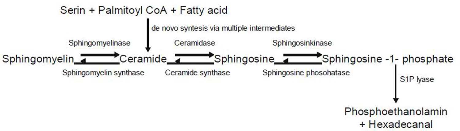
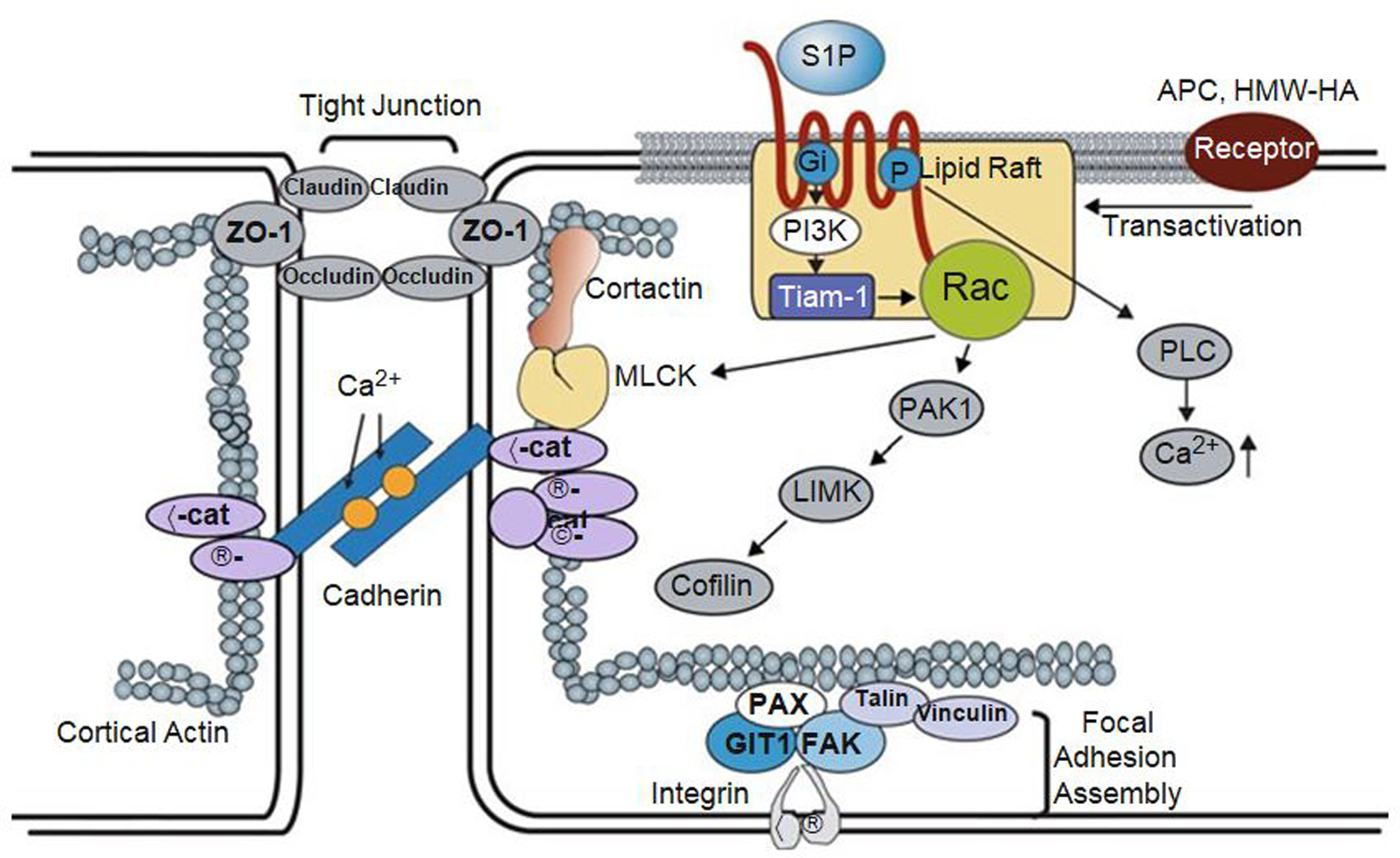
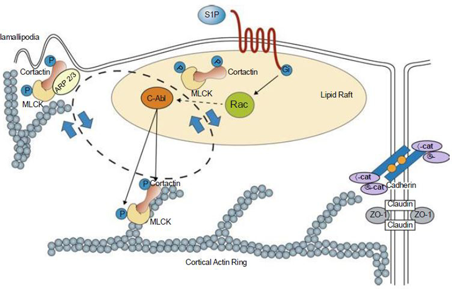
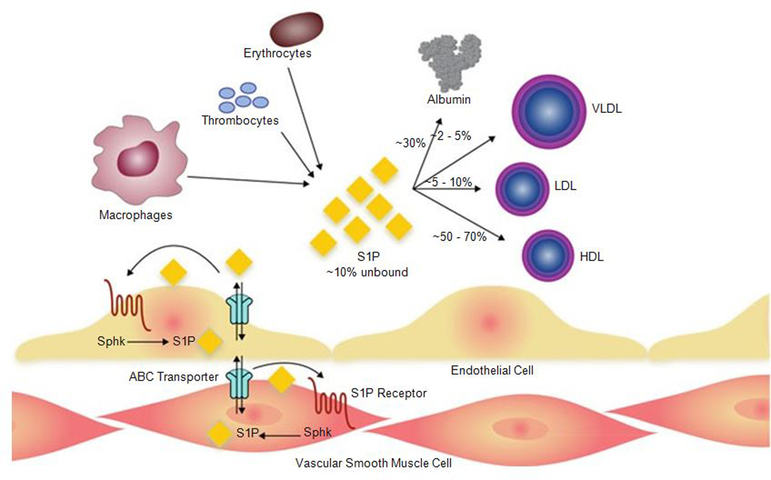
Of the multiple and diverse homeostatic events that involve the lung vascular endothelium, participation in preserving vascular integrity and therefore organ function is paramount. We were the first to show that the lipid growth factor and angiogenic factor, sphingosine-1-phosphate, is a critical agonist involved in regulation of human lung vascular barrier function (Garcia et al. J Clin Invest, 2011). Utilizing both in vitro models and preclinical murine, rat, and canine models of acute and chronic inflammatory lung injury, we have shown that S1Ps, as well as multiple S1P analogues such as FTY720 and ftysiponate, serve as protective agents limiting the disruption of the vascular EC monolayer in the pulmonary microcirculation and attenuate parenchymal accumulation of inflammatory cells and high protein containing extravasated fluid, thereby reducing interstitial and alveolar edema. The vasculo-protective mechanism of these therapeutic effects occurs via ligation of specific G-protein-coupled receptors and an intricate interplay of S1P with other factors (such as MAPKS, ROCKs, Rho, Rac1) with rearrangement of the endothelial cytoskeleton to form strong cortical actin rings in the cell periphery and enhanced cell-to-cell and cell-to-matrix tethering dynamics. This cascade leads to reinforcement of focal adhesions and paracellular junctional complexes via cadherin, paxillin, catenins, and zona occludens. S1P through its interaction with Rac and Rho influences the cytoskeletal rearrangement indicated in the later stages of angiogenesis as a stabilizing force, preventing excessive vascular permeability. These properties translate into a therapeutic potential for acute and chronic inflammatory lung injuries. S1P has potential for providing a paradigm shift in the approach to disruption of critical endothelial gatekeeper function, loss of lung vascular integrity, and increased vascular permeability, defining features of acute lung injury (ALI), and may prove to exhibit an intrinsically protective role in the pulmonary vasculature ameliorating agonist- or sepsis-induced pulmonary injury and vascular leakage.


Figures

Fig. 1
Fig. 1
Ceramide is either formed de novo from serine, palmitoyl coA, and fatty acid or via breakdown of membrane sphingomyelin. Ceramide is further converted to sphingosine, which can be phosphorylated to generate S1P. Degradation of S1P could be reversible by dephosphorylation or irreversible by S1P lyase. S1P, sphingosine-1-phosphate [Modified from Schuchardt et al. (2011)]
Fig. 2
Fig. 2
Regulation of vascular permeability by S1P/ SIP1 signaling. Binding of S1P to the SIP1 receptor stimulates the Gi-dependent recruitment of PI3 kinase, Tiam1, and Rac1 to lipid rafts (CEM), which serves to activate Rac1 in a Gi-PI3K-Tiam1-dependent manner. In addition, S1P induces an increase in intracellular Ca2+ concentration via a Gi-PLC pathway with additional activation of Rac1. After the activation of Rac1, S1P induces a series of profound events including adherens junction and tight junction assembly, cytoskeletal reorganization, and formation of focal adhesions that combine to enhance vascular barrier function. Furthermore, the transactivation of S1P1 signaling by other barrier-enhancing agents is recently recognized as a common mechanism for promoting endothelial barrier function. TJ tight junction, AJ adherens junction, S1P sphingosine-1-phosphate, SIP1 sphingosine-1-phosphate receptor 1, PI3K phosphoinositide 3-kinase, Tiam 1 t-lymphoma invasion and metastasis gene 1, Rac1 Rho family of GTPase Rac1, PAK1 p21-activated protein kinase 1, LIMK LIM kinase, PLC phospholipase C, ZO-1 zona occluden protein-1, nmMLCK non-muscle myosin light-chain kinase, VE-Cad vascular endothelial cadherin, a-Cat a-Catenin, β-Cat β-Catenin, Vin vinculin, Pax paxillin, FAK focal adhesion kinase, GIT2 G-protein-coupled receptor kinase interactor-1, ECM extracellular matrix, APC activated protein C, HMW-HA high molecular weight hyaluronan [Modified from Wang and Dudek (2009)]
Fig. 3
Fig. 3
S1P regulates enhanced EC barrier function. Ligation of the S1PR1 Gi protein-coupled receptor by S1P rapidly (within 1–5 min) activates Rac and recruits signaling molecules and cytoskeletal effectors such as c-Abl, cortactin, and nmMLCK to lipid rafts (or CEMs). Tyrosine phosphorylation of these molecules is observed both in lipid rafts and at the EC periphery in association with cortical actin and lamellipodia formation. This activated complex likely interacts with Arp 2/3 machinery to produce lamellipodia protrusion at the cell periphery, which serves to increase overlap between adjacent ECs. The initiation and precise sequence of events responsible for these protein movements are unclear, but within 5 min after S1P stimulation, these proteins are found simultaneously distributed in lipid rafts, cortical actin structures, and peripheral membrane ruffling/lamellipodia (indicated by the bidirectional circle). S1P also induces adherens junction (AJ) and tight junction (TJ) assembly that serve to further strengthen the endothelial barrier. Multiple other signaling and cytoskeletal effector molecules participate in this process as reviewed elsewhere (Wang and Dudek 2009). MLCK non-muscle myosin light-chain kinase, VE-cad vascular endothelial cadherin, ZO-1 zona occluden protein-1 [Modified from Belvitch and Dudek (2012)]
Fig. 4
Fig. 4
Secretion of S1P by erythrocytes, platelets, macrophages, and endothelium. Once secreted, most of the S1P is uptaken by serum albumin or various serum lipoproteins. Intracellular-produced S1P in ECs or vascular smooth muscle cells could be transported across the membrane by ABC transporters. HDL high-density lipoprotein, LDL low-density lipoprotein, Sphk sphingosine kinase, S1P sphingosine-1-phosphate, VLDL very low-density lipoprotein [Modified from Belvitch and Dudek (2012)]

References

    1. Adyshev DM, Moldobaeva NK, Elangovan VR, Garcia JG, Dudek SM (2011) Differential involvement of ezrin/radixin/moesin proteins in sphingosine 1-phosphate-induced human pulmonary endothelial cell barrier enhancement. Cell Signal 23(12):2086–2096 - PMC - PubMed
    1. Amano M, Ito M, Kimura K, Fukata Y, Chihara K, Nakano T, Matsuura Y, Kaibuchi K (1996) Phosphorylation and activation of myosin by Rho-associated kinase (Rho-kinase). J Biol Chem 271(34):20246–20249 - PubMed
    1. An S, Zheng Y, Bleu T (2000) Sphingosine 1-phosphate-induced cell proliferation, survival, and related signaling events mediated by G protein-coupled receptors Edg3 and Edg5. J Biol Chem 275(1):288–296 - PubMed
    1. Arce FT, Whitlock JL, Birukova AA, Birukov KG, Arnsdorf MF, Lal R, Garcia JG, Dudek SM (2008) Regulation of the micromechanical properties of pulmonary endothelium by S1P and thrombin: role of cortactin. Biophys J 95(2):886–894 - PMC - PubMed
    1. Argraves KM, Wilkerson BA, Argraves WS, Fleming PA, Obeid LM, Drake CJ (2004) Sphingosine-1-phosphate signaling promotes critical migratory events in vasculogenesis. J Biol Chem 279(48): 50580–50590 - PubMed

MeSH terms

LinkOut - more resources