Review
. 2013 Nov;70(21):4101-16.
doi: 10.1007/s00018-013-1329-x. Epub 2013 Apr 7.

Cadherin mechanotransduction in tissue remodeling

Affiliations
Review

Cadherin mechanotransduction in tissue remodeling

Floor Twiss et al. Cell Mol Life Sci. 2013 Nov.

Abstract

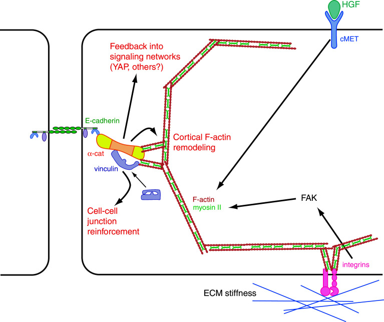
Mechanical forces are increasingly recognized as central factors in the regulation of tissue morphogenesis and homeostasis. Central to the transduction of mechanical information into biochemical signaling is the contractile actomyosin cytoskeleton. Fluctuations in actomyosin contraction are sensed by tension sensitive systems at the interface between actomyosin and cell adhesion complexes. We review the current knowledge about the mechanical coupling of cell-cell junctions to the cytoskeleton and highlight the central role of α-catenin in this linkage. We assemble current knowledge about α-catenin's regulation by tension and about its interactions with a diversity of proteins. We present a model in which α-catenin is a force-regulated platform for a machinery of proteins that orchestrates local cortical remodeling in response to force. Finally, we highlight recently described fundamental processes in tissue morphogenesis and argue where and how this α-catenin-dependent cadherin mechanotransduction may be involved.


Figures

Fig. 1
Fig. 1
Original description and definition of epithelial cell–cell junctions. An EM image from the original paper [1] that shows the zonula occludens (ZO) that contains the tight junctions, the zonula adherens (ZA) containing the adherens junctions and the macula adherens with the desmosomes (D) in the epithelium of the guinea pig
Fig. 2
Fig. 2
A working model to classify cadherin-containing adhesions and their transitions, based on their appearance in 2D cell culture systems. IF images and schematic representations of three different types of cadherin-based cell–cell adhesions. Myosin II dependent focal adherens junctions (FAJs) are formed upon initial cadherin–cadherin contacts, for instance in calcium-switch experiments in 2D cell culture. Tension-dependent presence of vinculin has been observed in these junctions. Myosin-based contractility in the radially oriented F-actin bundles and the intracellular network they emanate from is likely to determine the amount of tension, and concomitant stretched status of α-catenin, at the cadherin complex in FAJs. Maturation of these junctions results in the formation of linear adherens junctions (LAJs) that colocalize with thin F-actin structures and are aligned by thicker parallel F-actin bundles. Vinculin is largely absent from the cadherin complex in this conformation, which indicates that little tension is applied at the cadherin complex and α-catenin is in its unstretched state. LAJs can revert to FAJs in a myosin II-dependent manner during tissue remodeling processes such as endothelial permeabilization and epithelial cell scattering. LAJs further mature into zonula adherens junctions (ZAJs) when epithelial cells polarize and reorganize their cytoskeleton to form the apical F-actin belt that connects to the ZAJs. This process depends on myosin II activity as well as vinculin recruitment. The strong presence of vinculin in ZAJs may indicate high levels of tension and α-catenin stretching in the cadherin complex in this conformation, but the existence of a myosin II-independent pool of vinculin has also been reported
Fig. 3
Fig. 3
Trans-interactions between actin linkers of diverse cell–cell adhesion receptors. Schematic representation of the interconnectivity between actin linkers of the three main F-actin connected cell–cell adhesion complexes. α-catenin’s domains are labeled D1D5 according to their homology to its closest relative vinculin. The D2 domain is present in vinculin, but not in α-catenin. The D3 and D4 domains of α-catenin interact with afadin. The D4 domain alone can also interact, while the D3 domain alone does not [60]. The domain of interaction within afadin was not precisely mapped. Through its tail domain (D5), α-catenin interacts with the guanyl kinase (GUK) domain of ZO family proteins [64]. ZO proteins and afadin also interact with each other through their SH3 and proline-rich (PR) domains [62]
Fig. 4
Fig. 4
Structural and signaling role of α-catenin. The F-actin and β-catenin binding domains of α-catenin are critical for the structural support of cadherin-dependent cell–cell adhesions. For the support of basic cadherin adhesion, these domains can be replaced by unrelated peptide sequences that can bridge E-cadherin. For proper maturation of cell–cell junctions; and for their correct remodeling in tissues, the central domains of α-catenin are needed. In the current model, an intramolecular interaction between domains D4 and D3a is relieved when force across the cadherin-F-actin linkage increases. A binding site for vinculin, and likely additional proteins, is liberated in this way to induce the signals that drive adhesion maturation or junction remodeling. The proteins currently known to specifically interact with the central region of α-catenin are depicted. Of these, only the recruitment of vinculin has been shown to be myosin II-dependent
Fig. 5
Fig. 5
α-catenin as a platform for F-actin remodeling machinery. Many of α-catenin’s reported interactors (direct or indirect) can bind and/or regulate F-actin. This figure depicts these interacting proteins and classifies them into F-actin binders, F-actin remodelers, or proteins with a different function
Fig. 6
Fig. 6
Speculation on the role of cadherin mechanotransduction in junction deformation. As measured during Drosophila mesoderm invagination, apical constriction processes through pulsatile actomyosin contractions leading to deformation of cell–cell adhesions that are stabilized from relapse during the intermittent phases of low contractility. We speculate that increased tension on the cadherin complex, during the contractile peaks, results in α-catenin-dependent recruitment of an F-actin remodeling machinery that drive cortical remodeling to stabilize cell shape and maintain deformation in the relaxation phases. This could explain the ratchet-like manner in which pulsatile actomyosin contraction drives progressive shortening of cell–cell junctions in several developmental processes
Fig. 7
Fig. 7
Crosstalk between integrin and cadherin mechanotransduction in tissue remodeling. Induction of actomyosin contractility is a central event in crucial processes of tissue remodeling in vertebrates. HGF and other growth factors induce it during branching morphogenesis. ECM stiffness enhances it in tumor progression and stem cell differentiation. Integrin-based FAK activation is one of the key mechanisms by which integrin-mediated adhesion complexes control actomyosin. Increased contractility in the actomyosin network will lead to increased tension at sites of linkage to the cell cortex. At the cadherin–F-actin interface, increased tension will lead to allosteric activation of α-catenin and possibly other proteins. Based on current models, this may influence junctional strength through vinculin and cortical F-actin remodeling to the recruitment of several regulators. Feedback into more distant intracellular signaling networks is also expected, but the molecular details responsible have not been elucidated

References

    1. Farquhar MG, Palade GE. Junctional complexes in various epithelia. J Cell Biol. 1963;17:375–412. doi: 10.1083/jcb.17.2.375. - DOI - PMC - PubMed
    1. Volk T, Geiger B. A 135-kd membrane protein of intercellular adherens junctions. EMBO J. 1984;3(10):2249–2260. - PMC - PubMed
    1. Takeichi M. Functional correlation between cell adhesive properties and some cell surface proteins. J Cell Biol. 1977;75(2 Pt 1):464–474. doi: 10.1083/jcb.75.2.464. - DOI - PMC - PubMed
    1. Yoshida-Noro C, Suzuki N, Takeichi M. Molecular nature of the calcium-dependent cell–cell adhesion system in mouse teratocarcinoma and embryonic cells studied with a monoclonal antibody. Dev Biol. 1984;101(1):19–27. doi: 10.1016/0012-1606(84)90112-X. - DOI - PubMed
    1. Peyrieras N, et al. Uvomorulin: a nonintegral membrane protein of early mouse embryo. Proc Natl Acad Sci USA. 1983;80(20):6274–6277. doi: 10.1073/pnas.80.20.6274. - DOI - PMC - PubMed

Publication types

LinkOut - more resources