. 2013 May 24;288(21):14756-68.
doi: 10.1074/jbc.M113.466169. Epub 2013 Apr 5.

Minimal interleukin 6 (IL-6) receptor stalk composition for IL-6 receptor shedding and IL-6 classic signaling

Affiliations

Minimal interleukin 6 (IL-6) receptor stalk composition for IL-6 receptor shedding and IL-6 classic signaling

Paul Baran et al. J Biol Chem. .

Abstract

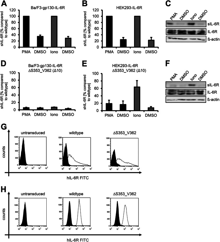
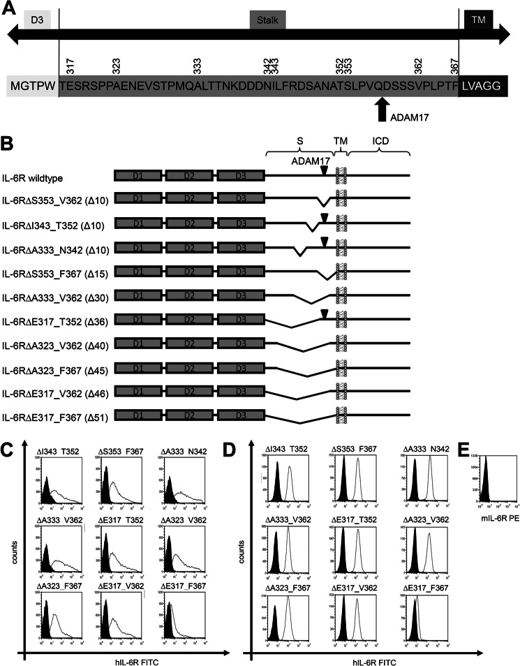
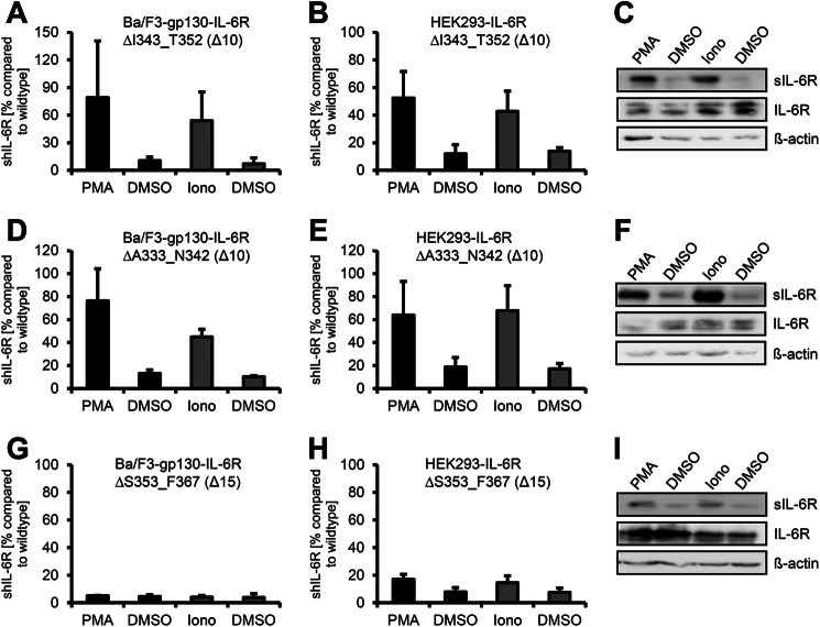
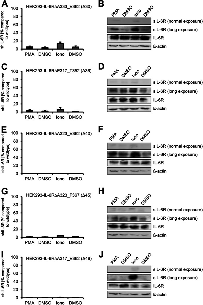
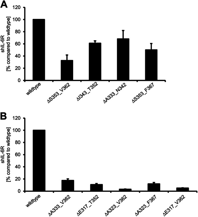
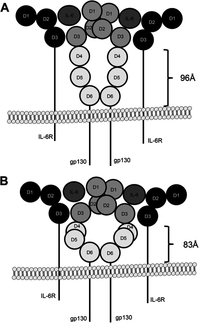
Signaling of the pleiotropic cytokine Interleukin-6 (IL-6) is coordinated by membrane-bound and soluble forms of the IL-6 receptor (IL-6R) in processes called classic and trans-signaling, respectively. The soluble IL-6R is mainly generated by ADAM10- and ADAM17-mediated ectodomain shedding. Little is known about the role of the 52-amino acid-residue-long IL-6R stalk region in shedding and signal transduction. Therefore, we generated and analyzed IL-6R stalk region deletion variants for cleavability and biological activity. Deletion of 10 amino acids of the stalk region surrounding the ADAM17 cleavage site substantially blocked IL-6R proteolysis by ADAM17 but only slightly affected proteolysis by ADAM10. Interestingly, additional deletion of the remaining five juxtamembrane-located amino acids also abrogated ADAM10-mediated IL-6R shedding. Larger deletions within the stalk region, that do not necessarily include the ADAM17 cleavage site, also reduced ADAM10 and ADAM17-mediated IL-6R shedding, questioning the importance of cleavage site recognition. Furthermore, we show that a 22-amino acid-long stalk region is minimally required for IL-6 classic signaling. The gp130 cytokine binding sites are separated from the plasma membrane by ~96 Å. 22 amino acid residues, however, span maximally 83.6 Å (3.8 Å/amino acid), indicating that the three juxtamembrane fibronectin domains of gp130 are not necessarily elongated but somehow flexed to allow IL-6 classic signaling. Our findings underline a dual role of the IL-6R stalk region in IL-6 signaling. In IL-6 trans-signaling, it regulates proper proteolysis by ADAM10 and ADAM17. In IL-6 classic-signaling, it acts as a spacer to ensure IL-6·IL-6R·gp130 signal complex formation.

Keywords: ADAM ADAMTS; ADAM10; ADAM17; IL-6 Signal Transduction; Interleukin; Interleukin-6; Interleukin-6 Receptor; Proteolytic Enzymes; Receptor Structure-Function; Signal Transduction.


Figures

FIGURE 1.
FIGURE 1.
ADAM17 and ADAM10 use different cleavage sites of the human IL-6R. A, Ba/F3-gp130-hIL-6R cells were treated for 2 h with PMA (100 nm) or for 1 h with ionomycin (Iono, 1 μm). Soluble IL-6R was measured by ELISA. The amount of soluble cytokine receptor without stimulation was considered as constitutive shedding and set to 1. Based on this, the increase of soluble receptors was calculated. B, HEK293 cells were transfected with an expression plasmid encoding wild-type human IL-6R. Cells were treated as described in panel A, sIL-6R was measured by ELISA, and values were calculated accordingly. C, transiently transfected HEK293 cells were treated as described under panel A. To determine the soluble cytokine receptors via Western blotting, they were precipitated from conditioned media with concanavalin A-covered Sepharose beads and visualized with 4-11 antibody (IL-6R). Cells were lysed after stimulation, and lysates were subsequently analyzed via Western blotting, whereas β-actin served as loading control. D, Ba/F3-gp130-IL-6RΔS353_V362 cells were treated as described in panel A. E and F, HEK293 cells were transfected with an expression plasmid encoding hIL-6RΔS353_V362. The experiment was performed as described in panels A and B. ELISA data are the mean (±S.D.) from three independent experiments, and Western blotting shows one representative experiment. G, cell surface expression of wild-type IL-6R and IL-6RΔS353_V362 on transiently transfected HEK293 cells was determined via flow cytometry as described under “Experimental Procedures.” H, cell surface expression on stably transduced Ba/F3-gp130-hIL-6R and Ba/F3-gp130-IL-6RΔS353_V362 cells was determined via flow cytometry as described under “Experimental Procedures.” One representative experiment of three performed is shown. The expressed IL-6R variant is given above the respective FACS plot.
FIGURE 2.
FIGURE 2.
Schematic overview of the human IL-6R constructs. A, shown is a schematic overview of the human IL-6R stalk region. The amino acid sequence of the human IL-6R from Met-311 to Gly-372 is shown. Amino acids belonging to the D3 region are shown in white, and amino acids belonging to the intracellular domain (ICD) are in dark gray. The stalk region is shown in light gray. Numbers above the sequence denote amino acids important for construction of deletion variants. The ADAM17 cleavage site is marked with a red arrow. B, the three extracellular domains of the human IL-6R are shown as dark gray-filled boxes (D1, D2, and D3). The other abbreviations used are stalk region (S), transmembrane region (TM), and intracellular domain (ICD). The known ADAM17 cleavage site Gln-357/ Asp-358 is marked with a black triangle in all constructs where this cleavage site is present. Deletions within the stalk regions are shown as kinks. The range of deleted amino acids is written in front of the schematic drawing, and the number of deleted amino acids given enclosed in parentheses. C, cell surface expression on transiently transfected HEK293 cells was determined via flow cytometry as described under “Experimental Procedures.” D, cell surface expression on stably transduced Ba/F3 cells was determined via flow cytometry as described under “Experimental Procedures.” E, the absence of endogenous IL-6R expression on Ba/F3-gp130 was stained with a murine IL-6R-specific antibody. One representative experiment of three performed is shown. The expressed IL-6R variant is given above the respective FACS plot. PE, phosphatidylethanolamine.
FIGURE 3.
FIGURE 3.
The ADAM10 cleavage site of the IL-6R is located close to the plasma membrane. A, Ba/F3-gp130-IL6R-ΔI343_T352 cells were treated for 2 h with PMA (100 nm) or for 1 h with ionomycin (Iono, 1 μm). Soluble IL-6R was measured by ELISA. The amount of soluble cytokine receptor without stimulation was considered as constitutive shedding and set to 1. Based on this, the increase of soluble receptors was calculated. B, HEK293 cells were transfected with an expression plasmid encoding IL-6RΔI343_T352. Cells were treated as described in panel A, sIL-6R was measured by ELISA, and values were calculated accordingly. C, transiently transfected HEK293 cells were treated as described in panel A. To determine the soluble cytokine receptors via Western blotting, they were precipitated from conditioned media with concanavalin A-covered Sepharose beads and visualized with 4–11 antibody (IL-6R). Cells were lysed after stimulation, and lysates were subsequently analyzed via Western blotting, whereas β-actin served as the loading control. D, Ba/F3-gp130-IL-6RΔA333_N342 cells were treated as described in panel A. E and F, HEK293 cells were transiently transfected with an expression plasmid encoding IL-6RΔA333_N342, and experiments were performed as described under panels (B and C). G, Ba/F3-gp130-IL-6RΔS353_F367 cells were treated as described in panel A. H and I, HEK293 cells were transiently transfected with an expression plasmid encoding IL-6RΔS353_F367, and experiments were performed as described in panels B and C.
FIGURE 4.
FIGURE 4.
Larger deletions within the stalk region abrogate ADAM10 and ADAM17 mediated shedding. HEK293 cells were transfected with expression plasmids encoding B IL-6RΔA333_V362 (A), IL-6RΔE317_T352 (C and D), IL-6RΔA323_V362 (E and F), IL-6RΔA323_F367 (G and H), or IL-6RΔE317_V362 (I and J). Soluble IL-6R was measured by ELISA. Precipitation and Western blotting was performed as described in the legend of Fig. 1. ELISA data are the mean ± S.D. from three independent experiments. Western blotting shows one representative experiment. Iono, ionomycin.
FIGURE 5.
FIGURE 5.
Deletions within the stalk region differentially influence constitutive IL-6R shedding. A and B, HEK293 cells were transfected with the indicated IL-6R constructs. 48 h after transfection medium was replaced with serum-free medium. The amount of constitutive shed IL-6R over a time period of 24 h was determined via ELISA. ELISA data are the mean ± S.D. from three independent experiments.
FIGURE 6.
FIGURE 6.
Influence of alterations of the stalk region on IL-6 dependent signal transduction. A, equal numbers of Ba/F3-gp130 cells were cultured for 2 days with increasing amounts of Hyper-IL-6 (0- 102 ng/ml) or human IL-6 (0- 102 ng/ml). RLU, relative light units. B–L, equal numbers of stably transduced Ba/F3-gp130 cells were cultured for 2 days with increasing amounts of Hyper-IL-6 (0–102 ng/ml) or human IL-6 (0–102 ng/ml). The IL-6R variant is given above the respective diagram. Cellular proliferation was quantified as indicated under “Experimental Procedures.” One representative experiment of two or three performed is shown.
FIGURE 7.
FIGURE 7.
Schematic drawing of two possible gp130 conformations. A, the three juxtamembrane fibronectin type III domains (D4-D6) of gp130 are in an elongated fashion, which means that the stalk of the IL-6R must be able to span a distance of at least 96 Å. B, in contrast, the structure published by Xu et al. (37) shows that domains D4-D6 of gp130 are not necessarily elongated but are somehow flexed, which would allow IL-6 classic signaling with a shorter stalk of about 83 Å.

References

    1. Scheller J., Chalaris A., Schmidt-Arras D., Rose-John S. (2011) The pro- and anti-inflammatory properties of the cytokine interleukin-6. Biochim. Biophys. Acta 1813, 878–888 - PubMed
    1. Garbers C., Hermanns H. M., Schaper F., Müller-Newen G., Grötzinger J., Rose-John S., Scheller J. (2012) Plasticity and cross-talk of interleukin 6-type cytokines. Cytokine Growth Factor Rev. 23, 85–97 - PubMed
    1. Sprecher C. A., Grant F. J., Baumgartner J. W., Presnell S. R., Schrader S. K., Yamagiwa T., Whitmore T. E., O'Hara P. J., Foster D. F. (1998) Cloning and characterization of a novel class I cytokine receptor. Biochem. Biophys. Res. Commun. 246, 82–90 - PubMed
    1. Boulanger M. J., Chow D. C., Brevnova E. E., Garcia K. C. (2003) Hexameric structure and assembly of the interleukin-6/IL-6 α-receptor/gp130 complex. Science 300, 2101–2104 - PubMed
    1. Jostock T., Müllberg J., Ozbek S., Atreya R., Blinn G., Voltz N., Fischer M., Neurath M. F., Rose-John S. (2001) Soluble gp130 is the natural inhibitor of soluble interleukin-6 receptor transsignaling responses. Eur. J. Biochem. 268, 160–167 - PubMed

MeSH terms