. 2013 Jun 7;288(23):16726-16737.
doi: 10.1074/jbc.M113.452540. Epub 2013 Apr 5.

Energetics and location of phosphoinositide binding in human Kir2.1 channels

Affiliations

Energetics and location of phosphoinositide binding in human Kir2.1 channels

Nazzareno D'Avanzo et al. J Biol Chem. .

Abstract

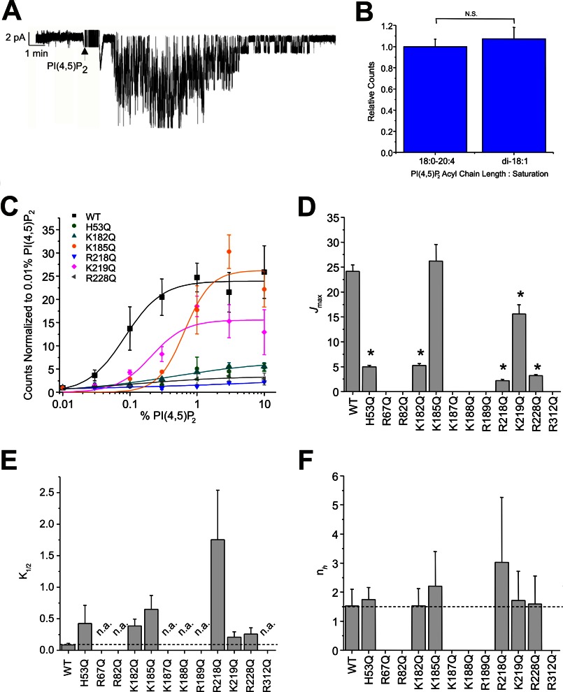
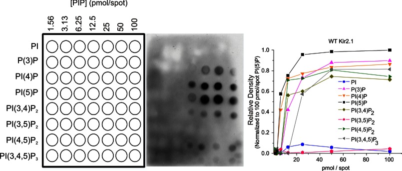
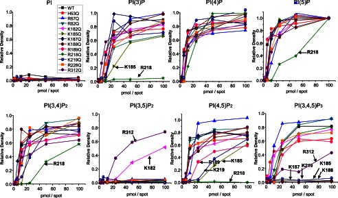
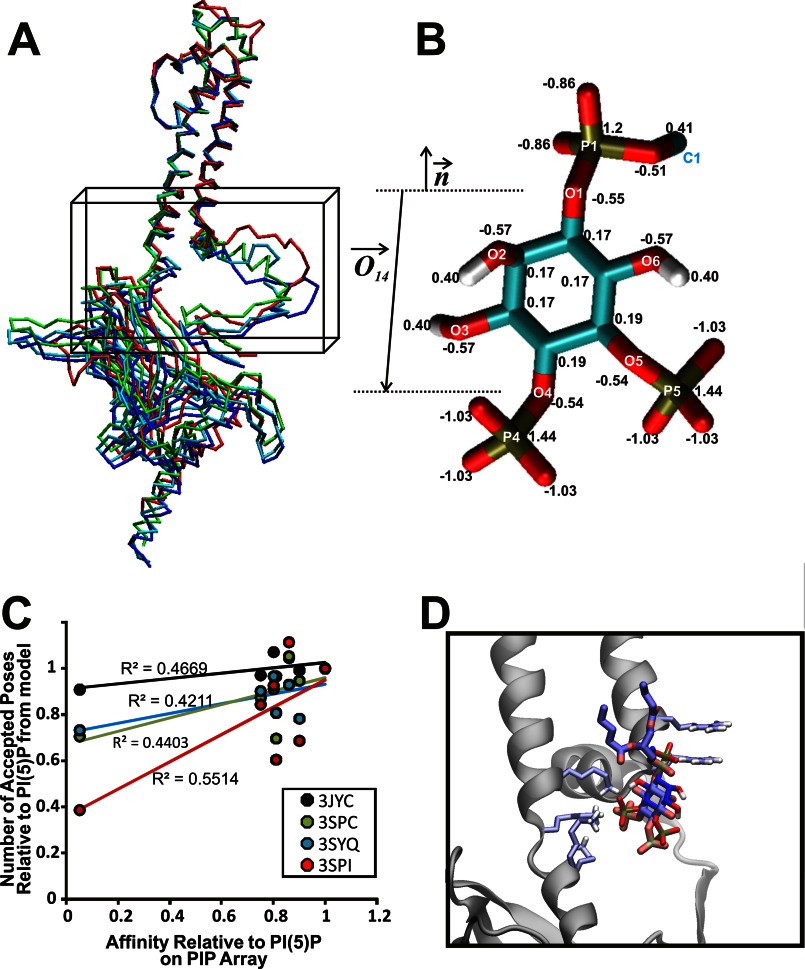
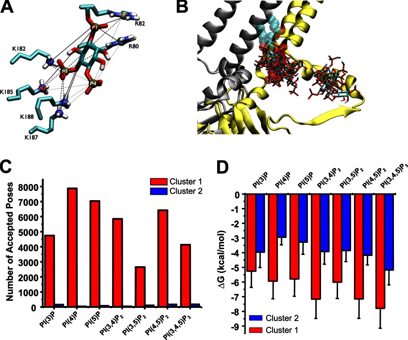
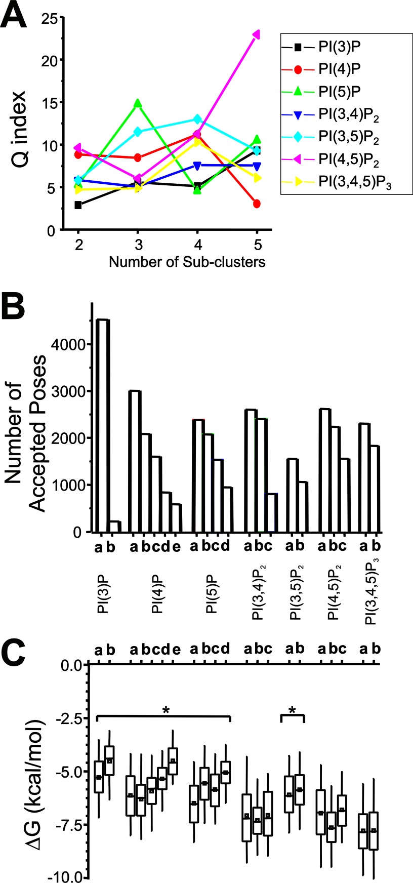
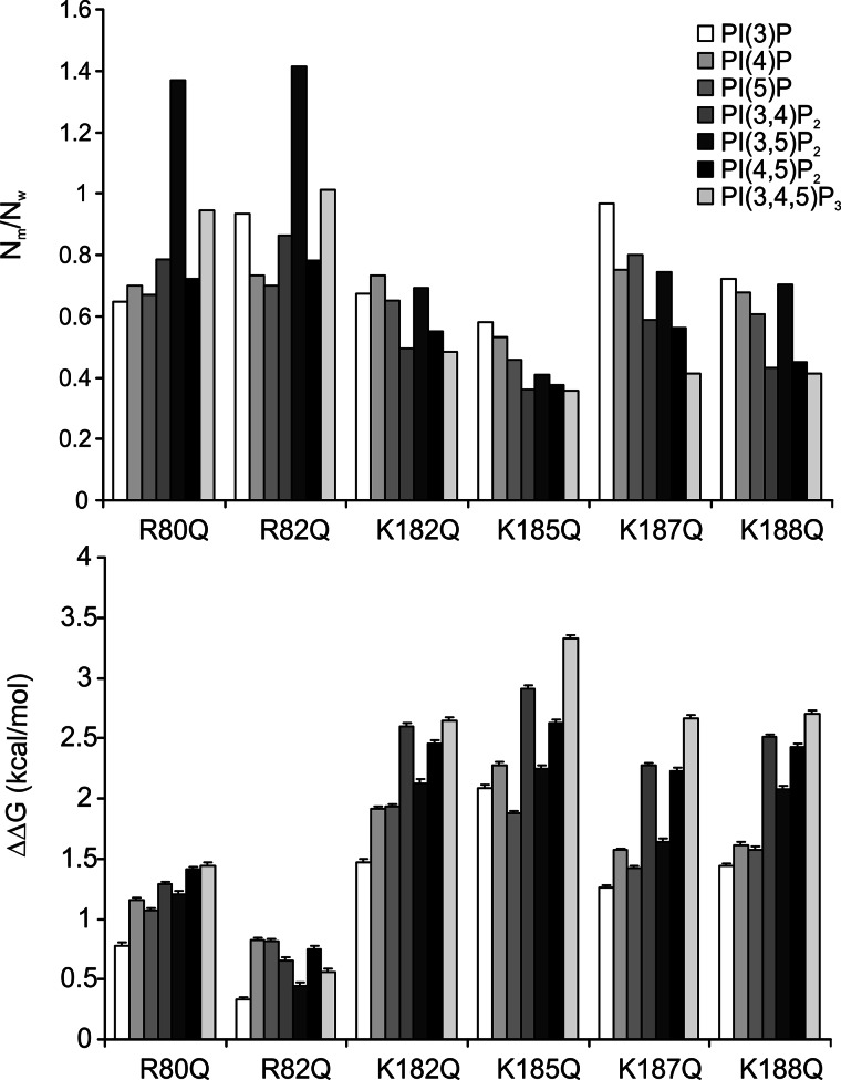
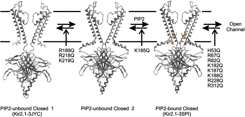
Kir2.1 channels are uniquely activated by phosphoinositide 4,5-bisphosphate (PI(4,5)P2) and can be inhibited by other phosphoinositides (PIPs). Using biochemical and computational approaches, we assess PIP-channel interactions and distinguish residues that are energetically critical for binding from those that alter PIP sensitivity by shifting the open-closed equilibrium. Intriguingly, binding of each PIP is disrupted by a different subset of mutations. In silico ligand docking indicates that PIPs bind to two sites. The second minor site may correspond to the secondary anionic phospholipid site required for channel activation. However, 96-99% of PIP binding localizes to the first cluster, which corresponds to the general PI(4,5)P2 binding location in recent Kir crystal structures. PIPs can encompass multiple orientations; each di- and triphosphorylated species binds with comparable energies and is favored over monophosphorylated PIPs. The data suggest that selective activation by PI(4,5)P2 involves orientational specificity and that other PIPs inhibit this activation through direct competition.

Keywords: Lipids; Molecular Docking; Molecular Modeling; Phosphatidylinositol Phosphatase; Potassium Channels.


Figures

FIGURE 1.
FIGURE 1.
A, superfusion of PI(4,5)P2 onto a patch of membrane containing Kir2.1 channels can recover channel activity after rundown. B, activation of Kir2.1 channels by PI(4,5)P2 is not sensitive to the acyl chain length or saturation as determined by comparing maximal activation between di-18:1 (dioleoyl) and 18:0–24:4 (stearoyl-arachidonoyl) PI(4,5)P2. C, PI(4,5)P2 concentration-activity relationship for WT and mutant Kir2.1 channels. For clarity, only mutants in which some activity could be observed are shown. Solid lines are fit curves using the Hill equation. No discernable activity was detected for R67Q, R82Q, K187Q, K188Q, R189Q, and R312Q Kir2.1 channels even in liposomes containing 30% PI(4,5)P2. D–F, parameters of fits to the Hill equation indicate that K185Q and K219Q mutants primarily shift the K1/2 of PI(4,5)P2, whereas H53Q, K182Q, R218Q, and R228Q mutations reduce the maximal flux as well as increase the K1/2 of PI(4,5)P2. No mutation significantly altered the Hill co-efficient (nH ≈ 1.5) which has been suggested to be a measure of co-operativity between ligands. Error bars, S.D.; N.S., not significant; n.a., no detectable activity.
FIGURE 2.
FIGURE 2.
The interaction of wild type Kir2.1 to PIP Arrays. A schematic diagram of PIP Arrays (left) shows an increasing amount of lipids from left to right. PIP Arrays were incubated with WT Kir2.1, and bound proteins were probed with an anti-His antibody (center). Densities in each array were internally normalized to density measured for the 100 pmol spot of PI(5)P (right).
FIGURE 3.
FIGURE 3.
The interaction of individual mutant Kir 2.1 to the PIP Arrays. Mutant Kir2.1 protein bound to the PIP Arrays was probed with an anti-His antibody. The raw images are shown in supplemental Fig. S2. Densitometry measurements from PIP Arrays were internally normalized to density measured for the 100 pmol spot of PI(5)P and are plotted for lipids individually. The mutations that caused >50% reduction in binding compared with WT Kir2.1 are designated by an arrow and residue name. These data indicate that for each PIP, it is a different subset of residues that when mutated to Gln (Q) disrupts channel binding. This suggests that these ligands orient differently in the binding pocket, thereby interacting with different subset of residues, which may explain why they do not equivalently trigger activation in Kir2.1 channels.
FIGURE 4.
FIGURE 4.
Docking of various PIPs to wild type Kir2.1 protein models. A, Cα trace of representative homology models of human Kir2.1 based on the chicken Kir2.2 structures (PDB ID codes 3JYC (blue), 3SPC (cyan), or 3SPI (green)) and mouse Kir3.2 (PDB ID code 3SYQ (red)). The search region for autodocking is designated by a black box. B, surrogate structure of the PI(4,5)P2 ligand with atom names and charges. The surrogates are the mimics of the PIPs with acyl chains cut off at the C1 position. Other PIPs were generated through substituting with either a hydroxyl or phosphate group at positions 3, 4, and 5 on the inositol ring. Atom charges are transferable to other PIPs. The vector for molecular axis (O⃗14) and bilayer normal (n⃗) are used for docking simulations and analysis. C, relative affinities (assessed by number of correctly oriented poses) for each ligand from our computational experiments compared with the relative binding affinities determined from the PIP Arrays. R2 values for the linear fit were 0.5514, 0.4669, 0.4403, and 0.4211 for 3SPI, 3JYC, 3SPC, and 3SYQ derived Kir2.1 models, respectively. This analysis suggests that the protein bound to the PIP Arrays most likely resides in a similar conformation to that observed in the closed PI(4,5)P2-bound structure (PDB ID code 3SPI), with weak binding in both cases to PI(3,5)P2, and so the remainder of our analysis was performed on docking simulations to models derived from this structure. D, docked pose of PI(4,5)P2 bound to Kir2.1–3SPI in darker color compared with the crystallographic PI(4,5)P2 bound to Kir2.2 in light color. Chicken Kir2.2 subunit A is shown schematically, and six basic residues in the binding pocket are shown as sticks.
FIGURE 5.
FIGURE 5.
A, distances between a ligand and the six putative binding residues. The pairs between the phosphorus atoms (P1, P4, and P5) of PI(4,5)P2 and the side chain atoms (CZ of Arg80, Arg82 and NZ of Lys182, Lys185, Lys187, and Lys188) are visualized by solid, dashed, and dotted lines. The distances of these pairs were used to cluster accepted poses. B, Kir 2.1–3SPI subunit A and D shown in yellow and silver ribbon, respectively, with the crystallographic PI(4,5)P2 bound to the subunit A shown in sphere translucently. An overlay of a single representative pose from each cluster of seven different C1-PIP ligands stacked on the Kir2.1–3SPI surface clearly indicates that these ligand bind primarily within two clusters. C, number of accepted poses within cluster 1 (red) and cluster 2 (blue) for each PIP. For each PIP, 96–99% of poses reside in cluster 1. D, average binding energy for poses in cluster 1 (red) and cluster 2 (blue) for each PIP docked to Kir2.1–3SPI.
FIGURE 6.
FIGURE 6.
A, the minimum value determined from the performance index (Q) versus cluster number taken to determine the optimal number of subclusters for PIP binding in cluster 1. B, the number of accepted poses in each subcluster for each of the seven PIP ligands docked to Kir2.1–3SPI. C, box-whisker plot of binding free energy (ΔGbinding in kcal/mol) within a subcluster for each PIP ligand. Monophosphorylated PIPs and PI(3,5)P2 bind with higher average energies than PI(4,5)P2; however, selective activation of Kir2.1 channels by PI(4,5)P2 cannot be solely accounted for based on this because PI(3,4)P2 and PI(3,4,5)P3 bind with similar energies.
FIGURE 7.
FIGURE 7.
Docking of various PIPs to Kir2.1 mutant protein models. The same docking simulations were carried out on the Kir2.1 model proteins with a single mutation as listed in this figure. Top, the number of accepted poses in cluster 1 relative to that of wild type protein is shown. Bottom, the difference of binding free energy in the cluster 1 between the wild type and each mutant protein is shown in kcal/mol unit. Error bars represent S.E.
FIGURE 8.
FIGURE 8.
Proposed model for the predominant pathway to channel activation. Kir2.1 channels may undergo a conformational change in which the cytoplasmic domain moves from a 3JYC type closed conformation (1) toward the plasma membrane and interacts through a hydrogen bond network with the slide helix, leading to a PI(4,5)P2 unbound structure (similar to what was observed for the Kir3.2 apo structure). We suggest that this transition may be less favored by mutations R189Q, R218Q, and K219Q, leading to reduced binding of PIP ligands. However, once the transition occurs, this state generates a three-dimensional binding pocket that strongly co-ordinates PI(4,5)P2, binding of which is disrupted by K185Q mutation. Further conformational changes lead to channel opening. Multiple mutations disrupt this transition, without affecting earlier steps of binding.

References

    1. Cheng W. W., Enkvetchakul D., Nichols C. G. (2009) KirBac1.1: it's an inward rectifying potassium channel. J. Gen. Physiol. 133, 295–305 - PMC - PubMed
    1. D'Avanzo N., Cheng W. W., Doyle D. A., Nichols C. G. (2010) Direct and specific activation of human inward rectifier K+ channels by membrane phosphatidylinositol 4,5-bisphosphate. J. Biol. Chem. 285, 37129–37132 - PMC - PubMed
    1. Enkvetchakul D., Jeliazkova I., Nichols C. G. (2005) Direct modulation of Kir channel gating by membrane phosphatidylinositol 4,5-bisphosphate. J. Biol. Chem. 280, 35785–35788 - PubMed
    1. Leal-Pinto E., Gómez-Llorente Y., Sundaram S., Tang Q. Y., Ivanova-Nikolova T., Mahajan R., Baki L., Zhang Z., Chavez J., Ubarretxena-Belandia I., Logothetis D. E. (2010) Gating of a G protein-sensitive mammalian Kir3.1 prokaryotic Kir channel chimera in planar lipid bilayers. J. Biol. Chem. 285, 39790–39800 - PMC - PubMed
    1. Cheng W. W., D'Avanzo N., Doyle D. A., Nichols C. G. (2011) Dual-mode phospholipid regulation of human inward rectifying potassium channels. Biophys. J. 100, 620–628 - PMC - PubMed

Publication types

MeSH terms

Substances