. 2013:Chapter 15:Unit 15.14.1-51.
doi: 10.1002/0471142735.im1514s101.

Autoimmune myocarditis, valvulitis, and cardiomyopathy

Affiliations

Autoimmune myocarditis, valvulitis, and cardiomyopathy

Jennifer M Myers et al. Curr Protoc Immunol. 2013.

Abstract

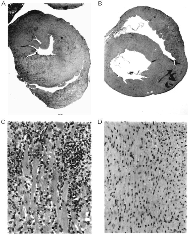
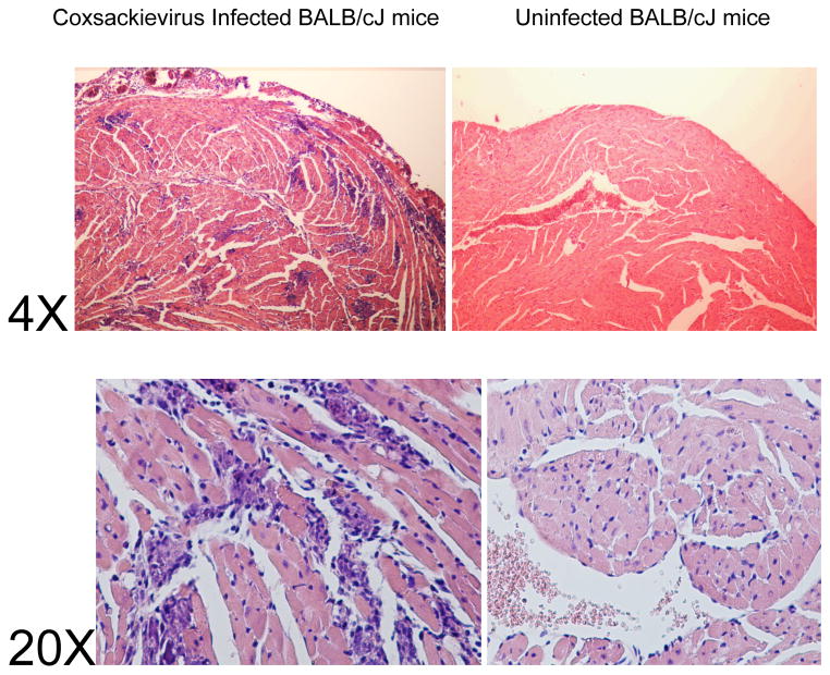
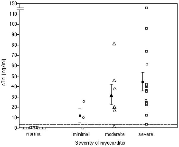
Myocarditis and valvulitis are inflammatory diseases affecting myocardium and valve. Myocarditis, a viral-induced disease of myocardium, may lead to dilated cardiomyopathy and loss of heart function. Valvulitis leads to deformed heart valves and altered blood flow in rheumatic heart disease. Animal models recapitulating these diseases are important in understanding the human condition. Cardiac myosin is a major autoantigen in heart, and antibodies and T cells to cardiac myosin are evident in inflammatory heart diseases. This unit is a practical guide to induction and evaluation of experimental autoimmune myocarditis (EAM) in several mouse strains and the Lewis rat. Purification protocols for cardiac myosin and protocols for induction of EAM by cardiac myosin and its myocarditis-producing peptides, and coxsackievirus CVB3, are defined. Protocols for assessment of myocarditis and valvulitis in humans and animal models provide methods to define functional autoantibodies targeting cardiac myosin, β-adrenergic, and muscarinic receptors, and their deposition in tissues.


Figures

Figure 15.14.1
Figure 15.14.1
Figure 15.14.1AHistopathology of EAM. Panels A and B are low-power (20×) and panels C and D are high-power (320×) photomicrographs of hematoxylin and eosin–stained cross-sections from hearts of A/J mice. Panels A and C demonstrate severe myocarditis in a mouse immunized with cardiac myosin. Panels B and D demonstrate uninflamed myocardium from a control mouse immunized with CFA only. (From Stacy Smith, MD and Paul Allen, PhD, Washington University, St Louis) Figure 15.14.1B. Myocarditis in male BALB/cJ mice infected with the H3 variant of CVB3. Male mice, 5–7 weeks of age were injected i.p. with 50 PFU H3 virus PBS or with PBS without virus (uninfected). Mice were killed 7 days after i.p. injection. Hearts were formalin fixed, paraffin embedded, sectioned and stained with hematoxylin and eosin. Histology is shown at 4X and 20X magnification. (From Sally Huber, PhD, University of Vermont, Colchester, VT)
Figure 15.14.1
Figure 15.14.1
Figure 15.14.1AHistopathology of EAM. Panels A and B are low-power (20×) and panels C and D are high-power (320×) photomicrographs of hematoxylin and eosin–stained cross-sections from hearts of A/J mice. Panels A and C demonstrate severe myocarditis in a mouse immunized with cardiac myosin. Panels B and D demonstrate uninflamed myocardium from a control mouse immunized with CFA only. (From Stacy Smith, MD and Paul Allen, PhD, Washington University, St Louis) Figure 15.14.1B. Myocarditis in male BALB/cJ mice infected with the H3 variant of CVB3. Male mice, 5–7 weeks of age were injected i.p. with 50 PFU H3 virus PBS or with PBS without virus (uninfected). Mice were killed 7 days after i.p. injection. Hearts were formalin fixed, paraffin embedded, sectioned and stained with hematoxylin and eosin. Histology is shown at 4X and 20X magnification. (From Sally Huber, PhD, University of Vermont, Colchester, VT)
Figure 15.14.2
Figure 15.14.2
cTnI values in murine EAM. Values represent cTnI levels measured 21 days after immunization with myosin/CFA or CFA alone (controls). The severity of myocarditis was determined histologically according to the grading scale in Table 15.14.4. Closed symbols represent the mean ± SE for each group. The dashed line represents the upper reference level for the assay. (From Stacy Smith, MD and Paul Allen, PhD, Washington University, St Louis, MO)

References

    1. Myocarditis and Giant Cell Myocarditis. from http://myocarditisfoundation.org.
    1. Abston ED, Barin JG, et al. IL-33 Independently Induces Eosinophilic Pericarditis and Cardiac Dilation: ST2 Improves Cardiac Function. Circ Heart Fail. 2012;5(3):366–375. - PMC - PubMed
    1. Abston ED, Coronado MJ, et al. Th2 regulation of viral myocarditis in mice: different roles for TLR3 versus TRIF in progression to chronic disease. Clin Dev Immunol. 2012;2012:129486. - PMC - PubMed
    1. Adams JE, 3rd, Abendschein DR, et al. Biochemical markers of myocardial injury. Is MB creatine kinase the choice for the 1990s? Circulation. 1993;88(2):750–763. - PubMed
    1. Afanasyeva M, Georgakopoulos D, et al. Impaired up-regulation of CD25 on CD4+ T cells in IFN-gamma knockout mice is associated with progression of myocarditis to heart failure. Proc Natl Acad Sci U S A. 2005;102(1):180–185. - PMC - PubMed

Publication types

MeSH terms

LinkOut - more resources