Review
. 2014 Jan;55(1):13-31.
doi: 10.1194/jlr.R031534. Epub 2013 Apr 6.

Cytochrome P450-mediated metabolism of vitamin D

Affiliations
Review

Cytochrome P450-mediated metabolism of vitamin D

Glenville Jones et al. J Lipid Res. 2014 Jan.

Abstract

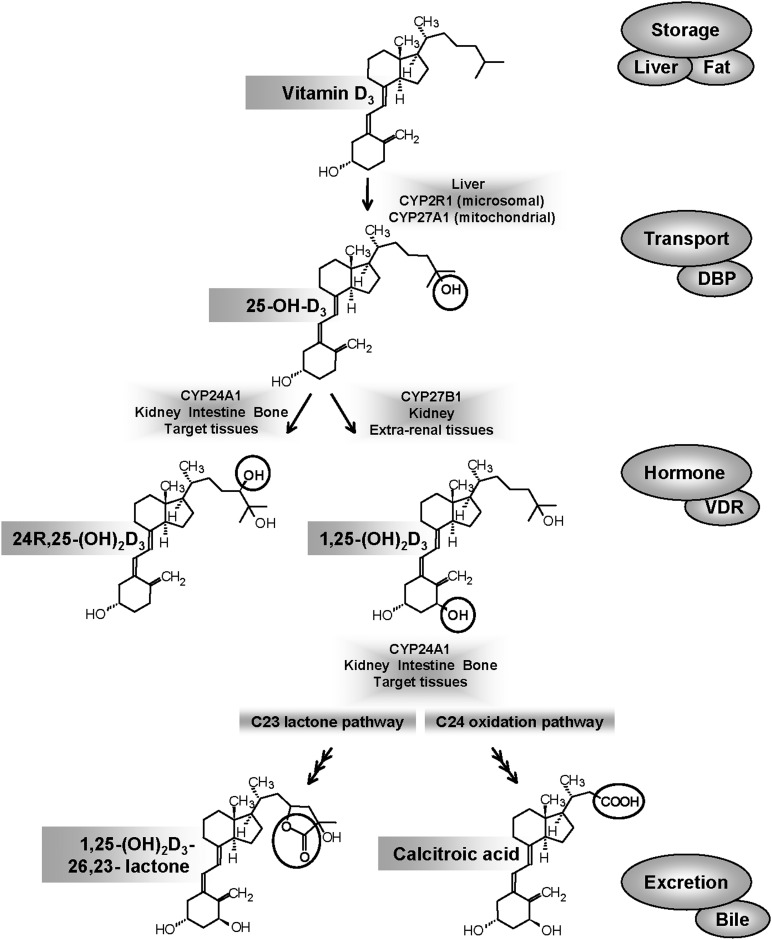
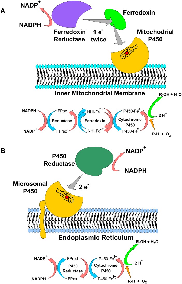
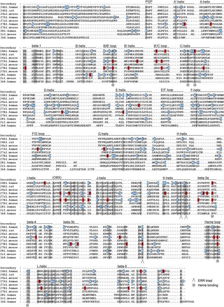
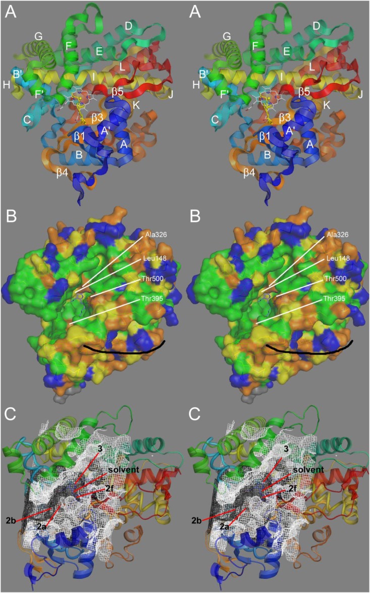
The vitamin D signal transduction system involves a series of cytochrome P450-containing sterol hydroxylases to generate and degrade the active hormone, 1α,25-dihydroxyvitamin D3, which serves as a ligand for the vitamin D receptor-mediated transcriptional gene expression described in companion articles in this review series. This review updates our current knowledge of the specific anabolic cytochrome P450s involved in 25- and 1α-hydroxylation, as well as the catabolic cytochrome P450 involved in 24- and 23-hydroxylation steps, which are believed to initiate inactivation of the vitamin D molecule. We focus on the biochemical properties of these enzymes; key residues in their active sites derived from crystal structures and mutagenesis studies; the physiological roles of these enzymes as determined by animal knockout studies and human genetic diseases; and the regulation of these different cytochrome P450s by extracellular ions and peptide modulators. We highlight the importance of these cytochrome P450s in the pathogenesis of kidney disease, metabolic bone disease, and hyperproliferative diseases, such as psoriasis and cancer; as well as explore potential future developments in the field.

Keywords: 1,25-(OH)2D3; CYP24A1; CYP27A1; CYP27B1; CYP2R1; chronic kidney disease; idiopathic infantile hypercalcemia; regioselectivity; vitamin D analog; vitamin D-dependent rickets.


Figures

Fig. 1.
Fig. 1.
Main cytochrome P450-mediated steps involved in vitamin D metabolism are depicted along with the main metabolites of vitamin D. Two other proteins, DBP and VDR, play essential roles in the transport of metabolites from one tissue to another and the key signal transduction events involved in target cell action, respectively.
Fig. 2.
Fig. 2.
Electron transport chains and protein components of the vitamin D hydroxylases. (A) In mitochondria, NADPH is oxidized by the flavoprotein ferredoxin reductase, which transfers single electrons through a pool of ferredoxin iron-sulfur proteins to the mitochondrial P450s on the inner membrane. (B) In the endoplasmic reticulum, electron equivalents from NADPH are captured by the NADPH P450 reductase (also known as P450 oxidoreductase, POR). The two electrons from NADPH are transferred sequentially to the microsomal P450 (e.g., CYP2R1). (Used from Ref. with permission.)
Fig. 3.
Fig. 3.
Sequence alignments of structurally determined or predicted secondary structures for vitamin D hydroxylases. Residues conserved in both mitochondrial and microsomal P450s (shaded) are structurally or functionally important, although elements of substrate recognition, binding, and specificity are inherently less conserved. Heme-binding residues and ERR-triad residues are also indicated. Locations of missense mutations leading to CYP2R1 deficiency (VDDR type 1B), CYP27A1 deficiency (CTX), CYP27B1 deficiency (VDDR type 1A), and CYP24A1 deficiency (IIH) are indicated by red shading. SNPs from dbSNP, Ensembl, Sanger Cosmic, and 1000 Genomes are shown in blue.
Fig. 4.
Fig. 4.
Structural analysis of CYP24A1. (A) Stereographic view of the CYP24A1 crystal structure (3k9v.pdb) with labeled secondary structures. An analysis of heme-ligand geometry in cytochrome P450s permitted docking of the substrate 1,25-(OH)2D3 (yellow) into the heme distal cavity active site. (B) Degree of amino-acid conservation in CYP24A1 observed across approximately 50 species orthologs: green (>95%), yellow (>85%), orange (>60%), and blue (<60%). The black curve indicates a possible membrane-binding surface. (C) Open active site cavity/cleft (white mesh) and an earlier model of a closed cavity (black mesh). Various access/egress channel trajectories are indicated. (Used with permission from Ref. 24).
Fig. 5.
Fig. 5.
Comparison of the enzymatic properties of two vitamin D-25-hydroxylases: CYP2R1 and CYP27A1. The substrates used are the prodrugs 1α-OH-D2 and 1α-OH-D3 to gauge the site and efficiency of the two vitamin D 25-hydroxylases toward D2 and D3 family members. Chromatograms of metabolites from in vitro reconstituted CYP2R1 enzyme (upper panels) (23) and transiently transfected CYP27A1 in COS-1 cells (lower panels) (39). Upper left and lower left panels represent traces from incubation with 1α-OH-D2. Upper right and lower right panels represent traces from incubation with 1α-OH-D3. The lack of CYP27A1-mediated 25-hydroxylation toward 1α-OH-D2 is evident, although 1α,24(OH)2D2 metabolite is detectable in the serum of animals given large doses of vitamin D2 (41) and is a VDR agonist.
Fig. 6.
Fig. 6.
(A) Physiological roles of renal CYP27B1 and CYP24A1 in calcium and phosphate homeostasis. Ca and PO4 ions through PTH, FGF-23, and the hormone 1,25-(OH)2D3 play key roles in determining the balance between the synthesis and degradation of 25-OH-D3 and 1α,25-(OH)2D3 by regulating renal CYP27B1 and CYP24A1, respectively. (B) Putative roles of extrarenal CYP27B1 and CYP24A1 in establishing the optimal target cell concentration of 1,25-(OH)2D3 for regulation of gene expression in noncalcemic functions. Cytokines are believed to regulate these extrarenal/target cell enzymes. Normal cells balance synthesis and degradation to generate optimal levels of 1,25-(OH)2D3. Cancer cells show reduced CYP27B1 and increased CYP24A1 expression. (Reproduced from Ref. 166)
Fig. 7.
Fig. 7.
Location of CYP24A1 polymorphisms (in SNP databases) and CYP24A1 missense mutations identified in patients with IIH. The relative positions of the conserved α-helices and β-strands in the CYP24A1 protein are indicated in yellow and blue, respectively. Secondary structures positioning substrate contact residues are located in the β-1, A-helix, B′-helix, B′/C-loop, F/G-loop, I-helix, β-3a, β-3b, and β-5 structures; heme-binding and ERR-triad residues stabilize protein structure and are involved in ferredoxin binding and electron transfer to the heme iron.

References

    1. DeLuca H. F. 1974. Vitamin D: the vitamin and the hormone. Fed. Proc. 33: 2211–2219 - PubMed
    1. Ponchon G., DeLuca H. F. 1969. The role of the liver in the metabolism of vitamin D. J. Clin. Invest. 48: 1273–1279 - PMC - PubMed
    1. Fraser D. R., Kodicek E. 1970. Unique biosynthesis by kidney of a biologically active vitamin D metabolite. Nature. 228: 764–766 - PubMed
    1. Hewison M., Adams J. S. 2011. Extrarenal 1α-hydroxylase. In Vitamin D. 3rd edition. D. Feldman, J. W. Pike, and J. S. Adams, editors. Academic Press, San Diego, CA. 777–806.
    1. Jones G., Strugnell S., DeLuca H. F. 1998. Current understanding of the molecular actions of vitamin D. Physiol. Rev. 78: 1193–1231 - PubMed

Supplementary concepts