. 2013;8(4):e57131.
doi: 10.1371/journal.pone.0057131. Epub 2013 Apr 2.

Global diversity of the Placozoa

Affiliations

Global diversity of the Placozoa

Michael Eitel et al. PLoS One. 2013.

Abstract

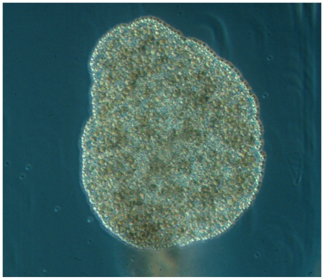
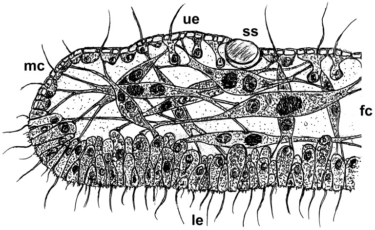
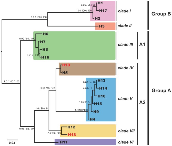
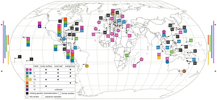
The enigmatic animal phylum Placozoa holds a key position in the metazoan Tree of Life. A simple bauplan makes it appear to be the most basal metazoan known and genetic evidence also points to a position close to the last common metazoan ancestor. Trichoplax adhaerens is the only formally described species in the phylum to date, making the Placozoa the only monotypic phylum in the animal kingdom. However, recent molecular genetic as well as morphological studies have identified a high level of diversity, and hence a potential high level of taxonomic diversity, within this phylum. Different taxa, possibly at different taxonomic levels, are awaiting description. In this review we firstly summarize knowledge on the morphology, phylogenetic position and ecology of the Placozoa. Secondly, we give an overview of placozoan morphological and genetic diversity and finally present an updated distribution of placozoan populations. We conclude that there is great potential and need to erect new taxa and to establish a firm system for this taxonomic tabula rasa.


Conflict of interest statement

Competing Interests: 1. The senior author Bernd Schierwater is a PLOS ONE Editorial Board member. This does not alter the authors' adherence to all the PLOS ONE policies on sharing data and materials. 2. Michael Eitel, Hans-Jürgen Osigus, and Bernd Schierwater are affiliated with the “Stiftung Tierärztliche Hochschule Hannover, ITZ, Ecology and Evolution, Hannover, Germany." The “Stiftung Tierärztliche Hochschule Hannover" is the German name for the “University of Veterinary Medicine, Hannover" and thus it is not a commercial company. The “ITZ, Ecology and Evolution" is a non-commercial research institute of this university.

Figures

Figure 1
Figure 1. Photograph of Trichoplax adhaerens, F.E. Schulze (1883).
The animal is about 0.5 mm in diameter. Photo by B. Schierwater, for additional images and videos of placozoan specimens see www.trichoplax.com and www.placozoa.de.
Figure 2
Figure 2. Revised schematic cross sections of a Placozoon.
Placozoans possess two epithelia, an upper epithelium facing the water and a lower epithelium facing the substrate. The traditional schematic cross section of a Placozoon shows a single fiber cell layer sandwiched between these layers. Recent studies by Guidi et al. , however, have shown that all morphologically studied genetic lineages, including the lineage originally used by Grell and Benwitz , actually possess several fiber cell layers. The given drawing thus represents the revised cross section of a typical placozoan specimen. It was also previously shown that the small marginal cells might represent the formerly described pluripotent stem cells . ue = upper epithelium; le = lower epithelium; fc = fiber cells; mc = marginal cells; ss = shiny sphere.
Figure 3
Figure 3. Maximum likelihood phylogenetic tree of metazoan relationships using a concatenated data matrix.
In this tree Placozoa group basal within the diploblasts (like in many other studies) but diploblasts and triploblasts occur as sister groups, a hypothesis that fits many morphological and structural genetics characters but is highly controversially discussed. From Schierwater et al., 2009 .
Figure 4
Figure 4. Unexpected diversity has been found in the phylum Placozoa.
Shown is the 16S Bayesian inference phylogram of different placozoan haplotypes. Presently seven genetically highly different clades (I–VII) have been identified. Haplotypes H18 and H19 have been newly identified in this study (red). Please note that the lineages H5 and H19 differ in just one nucleotide position within a highly variable loop region, which has been removed from the alignment (the result is the shown polytomy). Current knowledge on placozoan biodiversity is still limited and more samples are urgently needed. For details on phylogenetic analyses see Material and Methods and . Numbers beside nodes are from left to right: Bayesian posterior probabilities, Maximum likelihood and Maximum Parsimony bootstrap support. Values below 70% are marked with ‘-’.
Figure 5
Figure 5. Worldwide distribution of genetically characterized placozoan specimens.
The ten newly genotyped sites from this study are marked as circles and known genotypes from other studies are marked as squares. Samples from aquaria or holding tanks (‘aquarium samples’) with only a presumed/guessed origin are labeled with dashed lines. Note that several numbers combine multiple sampling sites. The color-coded bars and spots on the left and right mark the latitudinal distribution of the clades with at least two isolates from different locations each. Clades with a higher latitudinal distribution are also found in more habitats (see legend inlet). Modified from Eitel & Schierwater, 2010 . 1. Western Samoa; 2. Moorea, French Polynesia; 3. Oahu, Hawaii, US; 4. Monterey Bay, California, US (A.s.); 5. Pacific coast of Panama; 6. Twin Cays, Belize; 7. Puerto Morelos, Mexico and Roatan, Honduras; 8. Bahamas; 9. Cahuita, Costa Rica; 10. Galeta, Panama; 11. Discovery Bay, Jamaica; 12. Bocas del Toro, Panama; 13. Cubagua, Venezuela; 14. Grenada; 15. Puerto Rico; 16. Florida, US; 17. North Carolina, US; 18. Philadelphia, US; 19. Woods Hole, US; 20. Bermuda, GB; 21. Sao Sebastiao Channel, Brazil; 22. Puerto de la Cruz, Tenerife, Spain; 23. Quinta do Lorde Marina, Madeira, Portugal; 24. Roscoff, France; 25. Plymouth, GB (A.s.); 26. Banyuls-sur-Mer, France; 27. Castiglioncello, Italy; 28. Orbetello Lagoon, Italy; 29. Gulf of Trieste and Gulf of Naples, Italy; 30. Otranto, Italy; 31. Katerini and Ormos Panagias, Greece; 32. Turunc, Turkey; 33. Caesarea, Israel; 34. Almunecar, Granada, Spain; 35. Cala Rajada, Majorca, Spain ; 36. Cassis, France; 37. Yasmine and Zarzis, Tunisia; 38. San Felice Circeo, Italy; 39. Porto Cesareo, Italy; 40. Elat, Israel; 41. Mombasa, Kenya; 42. Reunion ; 43. Mauritius, France; 44. Laem Pakarang, Thailand; 45. Indonesia (A.s.); 46. Bali, Indonesia; 47. Adelaide, Australia; 48. ‘Indo-Pacific’ (A.s.); 49. Kota Kinabalu, Sabah, Malaysia ; 50. Zambales, Philippines; 51. Hong Kong, China; 52. Russian coast of the Sea of Japan; 53. Oki Island, Japan; 54. Shimoda, Honshu, Japan; 55. Shirahama, Honshu, Japan; 56. Chatan, Okinawa, Japan; 57. Iriomote, Ryukyu Islands, Japan; 58. Boracay, Philippines; 59. Guam, US; 60. Palau; 61. Manado, Sulawesi, Indonesia; 62. Madang, Papua New Guinea; 63. Lizard Island, Australia; 64. Townsville, Orpheus Island and Heron Island, Australia.
Figure 6
Figure 6. Coleman rarefaction curve.
The total number of different haplotypes is plotted against the number of genetically screened locations. A slight saturation effect is apparent starting at seven locations and eight haplotypes. The slope of the curve is, however, artificially reduced as continuously re-sampling of the two most frequent lineages occurs (H2 and H4). Both haplotypes together have been found in 47% of all genetically characterized locations. Extrapolation of the curve estimates the total number of placozoan haplotypes (species) to be higher than some 200.

References

    1. Appeltans W, Bouchet P, Boxshall GA, Fauchald K, Gordon DP, et al.. (2011) World Register of Marine Species. Accessed at http://wwwmarinespeciesorg on 2011-08-08.
    1. Schulze FE (1883) Trichoplax adhaerens, nov. gen., nov. spec. Zoologischer Anzeiger 6: 92–97.
    1. Schulze FE (1891) Über Trichoplax adhaerens. In: Reimer G, editor. Abhandlungen der Königlichen Preuss Akademie der Wissenschaften zu Berlin. Berlin: Verlag der königlichen Akademie der Wissenschaften. pp. 1–23.
    1. Behrendt G, Ruthmann A (1986) The cytoskeleton of the fiber cells of Trichoplax adhaerens (Placozoa). Zoomorphology 106: 123–130.
    1. Buchholz K, Ruthmann A (1995) The mesenchyme-like layer of the fibre cells of Trichoplax adhaerens: A syncytium. Zeitschrift für Naturforschung C 50c: 282–285. - PubMed

Publication types

LinkOut - more resources