. 2013;8(4):e60184.
doi: 10.1371/journal.pone.0060184. Epub 2013 Apr 2.

P2X7 integrates PI3K/AKT and AMPK-PRAS40-mTOR signaling pathways to mediate tumor cell death

Affiliations

P2X7 integrates PI3K/AKT and AMPK-PRAS40-mTOR signaling pathways to mediate tumor cell death

Shu Bian et al. PLoS One. 2013.

Abstract

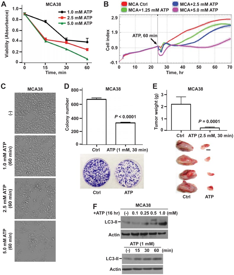
Background: Extracellular adenosine triphosphate (ATP) functions as a novel danger signal that boosts antitumor immunity and can also directly kill tumor cells. We have previously reported that chronic exposure of tumor cells to ATP provokes P2X7-mediated tumor cell death, by as yet incompletely defined molecular mechanisms.

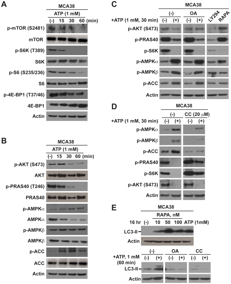
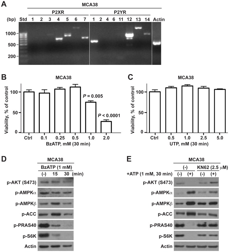
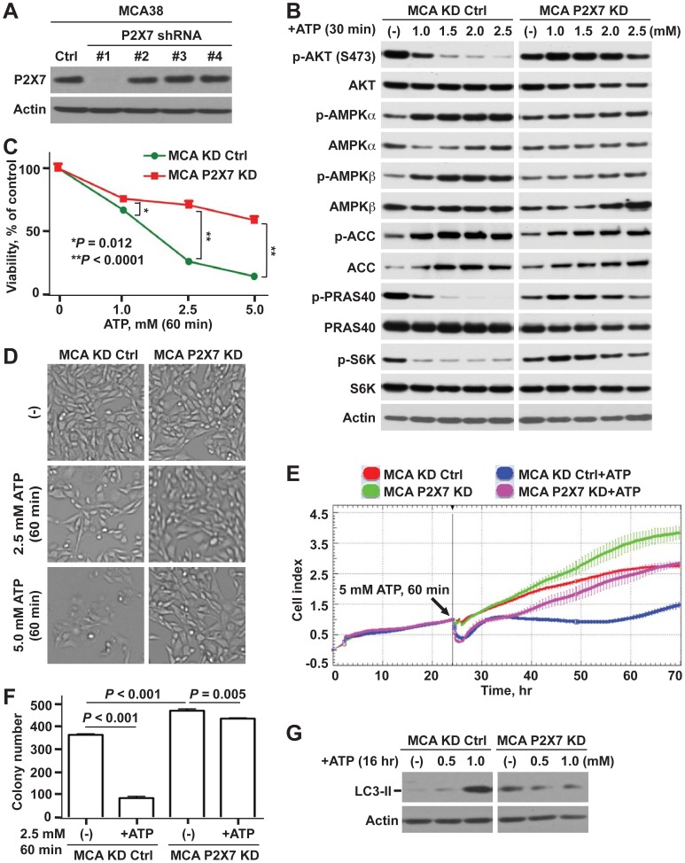
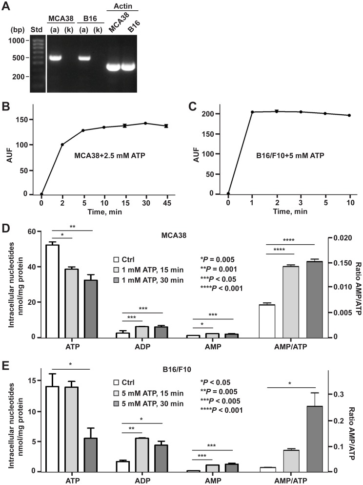
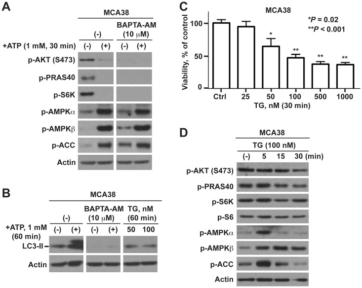
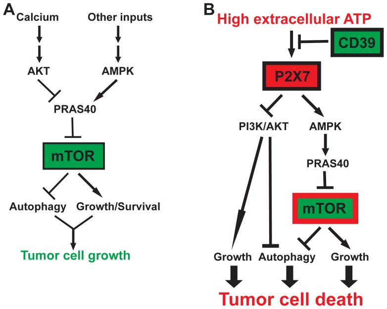
Methodology/principal findings: Here, we show that acute exposure of tumor cells to ATP results in rapid cytotoxic effects impacting several aspects of cell growth/survival, leading to inhibition of tumor growth in vitro and in vivo. Using agonist and antagonist studies together with generation of P2X7 deficient tumor cell lines by lentiviral shRNA delivery system, we confirm P2X7 to be the central control node transmitting extracellular ATP signals. We identify that downstream intracellular signaling regulatory networks implicate two signaling pathways: the known P2X7-PI3K/AKT axis and remarkably a novel P2X7-AMPK-PRAS40-mTOR axis. When exposed to high levels of extracellular ATP, these two signaling axes perturb the balance between growth and autophagy, thereby promoting tumor cell death.

Conclusions: Our study defines novel molecular mechanisms underpinning the antitumor actions of P2X7 and provides a further rationale for purine-based drugs in targeted cancer therapy.


Conflict of interest statement

Competing Interests: The authors have declared that no competing interests exist.

Figures

Figure 1
Figure 1. Antitumor effects of ATP on MCA38 colon cancer cells: in vitro and in vivo studies. A–C)
Dose- and time-dependent responses of MCA38 cells to ATP cytotoxicity at 24 hr after treatment: cell viability/proliferation by Cell Counting Kit-8 (CCK-8) (A); real-time and dynamic monitoring of cell growth by xCELLigence instrument (B); and representative bright field images of live cells by Celligo Cell Counting application (C). D) Top, colony numbers of MCA38 cells on day 12 post ATP treatment. Bottom, representative images of clonogenic assay dishes. E) Top, tumor weight of C57BL/6 mice implanted with untreated (Ctrl) or ATP-treated MCA38 cancer cells on day 14 (n = 12 per group). Bottom, representative tumors on day 14. Scale bar, 0.5 cm. F) Western blots of LC3-II, a sensitive indicator for autophagy, using MCA38 cells treated with ATP at various doses and times as indicated. β-actin serves as a loading control. Error bars, mean ± SEM. Data represent three to six experiments.
Figure 2
Figure 2. Intracellular mTOR, AKT, and AMPK signaling and induction of autophagy are impacted by cytotoxic ATP. A–B)
Western blot analysis of signaling components of mTOR (A), AKT and AMPK (B), in MCA38 cells after ATP treatment at different times as indicated. Inhibition of mTOR and AKT with concurrent stimulation of AMPK signaling was noted. C–D) Delineation of ATP-elicited intracellular signaling cascades by blockade of certain component using specific inhibitors, determined by Western blotting. OA (okadaic acid): 100 nM; LY294 (LY294002): 50 µM; RAPA (rapamycin): 100 nM; CC (compound C): 20 µM. E) Impacts of pathway inhibitors on autophagy in MCA38 cells, as examined by Western blots of LC3-II. β-actin is the loading control. Data represent three to six experiments.
Figure 3
Figure 3. ATP-induced tumor cell death is modulated via P2X7-mTOR axis.
A) Reverse transcription-PCR (RT-PCR) analysis of mRNA expression of P2 receptors in MCA38 cells. Size standards (Std) are shown in the left lane. B–C) Cell viability of MCA38 cells at 24 hr post treatment with BzATP (B) or UTP (C), as determined by CCK-8. Data are expressed as a percentage of untreated controls. D) Impacts of BzATP on AKT, AMPK, and mTOR signaling, as analyzed by Western blot. E) Blockade of P2X7 by antagonist KN62 abrogated ATP-induced inhibition on mTOR signaling in MCA38 cells, as evaluated by Western blotting. β-actin is shown as a loading control. Error bars, mean ± SEM. Data represent three to four experiments.
Figure 4
Figure 4. Knockdown of P2X7 decreases antitumor activity of ATP.
A) Western blots for P2X7 using lysates from MCA38 cells after knocking down P2X7 using lentiviral shRNAs. B–G) Differential effects of ATP on control knockdown (KD Ctrl) and P2X7 deficient (P2X7 KD) MCA38 cells: AKT- and AMPK-mTOR signaling by Western blotting (B); cell viability by CCK-8 (C); representative live cell images by Celligo (D); real-time measurement of cell growth by xCELLigence (E); clonogenicity by colony formation assay (F); and autophagy by Western blotting (G). β-actin is a loading control. Error bars, mean ± SEM. Data represent three experiments.
Figure 5
Figure 5. Characterization of P2X7 receptor function activated by tumoricidal ATP.
A) Examination of P2X7(a) or P2X7(k) variant expression in MCA38 and B16/F10 cells by RT-PCR with specific primer pairs for P2X7(a) or P2X7(k). Size standards (Std) are shown in the left lane. B–C) ATP-stimulated ethidium bromide uptake in MCA38 (B) and B16/F10 cells (C). D–E) Effects of extracellular ATP on intracellular levels of ATP, ADP and AMP in MCA38 (D) and B16/F10 cells (E) were measured by HPLC. Values of MCA38 AMP concentrations are multiplied five fold for the sole convenience of plotting on the same axes. Error bars, mean ± SEM. Data represent three experiments.
Figure 6
Figure 6. ATP-P2X7-mTOR elicited tumor cell death does not involve calcium signaling.
A) Effects of BAPTA-AM on ATP/P2X7-initiated AKT, AMPK and mTOR signaling in MCA38 cells, as examined by Western blotting. B) Effects of BAPTA-AM and thapsigargin (TG) on autophagy as determined by Western blots of LC3-II. C–D) Impacts of thapsigargin (TG) on MCA38 cell growth by CCK-8 (C); and Akt, AMPK and mTOR signaling by Western blot analysis (D). β-actin is the loading control. Error bars, mean ± SEM. Data represent three experiments.
Figure 7
Figure 7. Schematic illustration of how PI3K/AKT, AMPK and mTOR regulatory networks distinctly control tumor cell growth in response to high levels of ATP.
A) In the presence or absence of serum, tumor cell growth is dictated by cellular autophagy and growth/survival signals that are tightly controlled by PI3K/AKT and AMPK pathways convergent on mTOR by way of PRAS40. Physiological Ca2+ stores are absolutely required to maintain basal activation of the PI3K/AKT-PRAS40-mTOR signaling and cell growth. B) In a tumor microenvironment with pericellular ATP likely to be at high levels, activation of P2X7 on tumor cells leads to concurrent blockade of the mTOR signaling by way of AMPK-PRAS40 axis and the PI3K/AKT pathway. These are the two critical control nexuses for autophagy induction and growth inhibition that ultimately lead to tumor cell death. Ectonucleotidase CD39 protects tumor cells from antitumor activity of high levels of extracellular ATP , . Pathway key: →: Stimulation; –ı: Inhibition.

References

    1. White N, Burnstock G (2006) P2 receptors and cancer. Trends Pharmacol Sci 27: 211–217. - PubMed
    1. Deaglio S, Robson SC (2011) Ectonucleotidases as regulators of purinergic signaling in thrombosis, inflammation, and immunity. Adv Pharmacol 61: 301–332. - PMC - PubMed
    1. Eltzschig H, Sitkovsky M, Robson SC (2012) Purinergic signaling during Inflammation. N Engl J Med In press. - PubMed
    1. Volonte C, Amadio S, D’Ambrosi N, Colpi M, Burnstock G (2006) P2 receptor web: complexity and fine-tuning. Pharmacol Ther 112: 264–280. - PubMed
    1. Khakh BS, North RA (2006) P2X receptors as cell-surface ATP sensors in health and disease. Nature 442: 527–532. - PubMed

Publication types

MeSH terms

Substances

LinkOut - more resources