. 2013;8(4):e60469.
doi: 10.1371/journal.pone.0060469. Epub 2013 Apr 2.

The calpain/calpastatin system has opposing roles in growth and metastatic dissemination of melanoma

Affiliations

The calpain/calpastatin system has opposing roles in growth and metastatic dissemination of melanoma

Quentin Raimbourg et al. PLoS One. 2013.

Abstract

Conventional calpains are ubiquitous cysteine proteases whose activity is promoted by calcium signaling and specifically limited by calpastatin. Calpain expression has been shown to be increased in human malignant cells, but the contribution of the calpain/calpastatin system in tumorigenesis remains unclear. It may play an important role in tumor cells themselves (cell growth, migration, and a contrario cell death) and/or in tumor niche (tissue infiltration by immune cells, neo-angiogenesis). In this study, we have used a mouse model of melanoma as a tool to gain further understanding of the role of calpains in tumor progression. To determine the respective importance of each target, we overexpressed calpastatin in tumor and/or host in isolation. Our data demonstrate that calpain inhibition in both tumor and host blunts tumor growth, while paradoxically increasing metastatic dissemination to regional lymph nodes. Specifically, calpain inhibition in melanoma cells limits tumor growth in vitro and in vivo but increases dissemination by amplifying cell resistance to apoptosis and accelerating migration process. Meanwhile, calpain inhibition restricted to host cells blunts tumor infiltration by immune cells and angiogenesis required for antitumor immunity, allowing tumor cells to escape tumor niche and disseminate. The development of highly specific calpain inhibitors with potential medical applications in cancer should take into account the opposing roles of the calpain/calpastatin system in initial tumor growth and subsequent metastatic dissemination.


Conflict of interest statement

Competing Interests: The authors have declared that no competing interests exist.

Figures

Figure 1
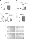
Figure 1. Calpain isoforms and calpastatin transgenic expression in B16-F10 cell line in culture.
A. Calpain isoform transcripts were quantified by PCR. The most abundant was m calpain (Cpn2) followed by µ calpain transcript (Cpn1) and their common sub-unit calpain 4 transcript (Cpns1). Calpastatin transcript (Cast1) was also detected. Means and error bars correspond to 4 independent RNA extracts. Gapdh was the housekeeping gene used for normalization. B. Calpastatin rabbit transgene expression was assessed by quantitative PCR. Calpastatin transgene was stably and efficiently expressed in B16-F10 clone (Calpast-Mel) but not in control B16-F10 clones (Ctrl-Mel). C. Calpastatin expression was assessed by Western Blot. Calpastatin protein was overexpressed in Calpast-Mel clones. D and E. Spectrin intact form and calpain-dependent breakdown products were assessed by Western Blot and optical density measure (OD). Calpastatin overexpression (Calpast-Mel) decreased spectrin degradation by calpains. N = 4 independent experiments, * p<0.05. F. Calpain activity in vitro was measured by specific substrate fluorescence. Calpain activity was decreased by calpastatin transgene. N = 6 independent experiments, * p<0.05.
Figure 2
Figure 2. Inhibition of calpains in both host and melanoma cells (global inhibition).
C57BL/6 control (WT mice) and transgenic mice (CalpTG mice) were injected with one million melanoma B16-F10 cells transfected with control plasmid (WT/Ctrl-Mel) and calpastatin plasmid (TG/Calpast-Mel) respectively. Mice were sacrificed at day 16 for tissue analysis. A. Tumor growth was measured from day 9 to day 16. Calpastatin overexpression in both hosts and melanoma decreased significantly tumor growth. N = 10/group, * p<0.05. B. Tumor weight at day 16. Calpastatin overexpression in both hosts and melanoma decreased non-significantly tumor weight. N = 10/group, p = NS. C. Angiogenesis assessed by vessel count at 200×magnification after CD-31 staining. TG/Calpast-Mel mice had a trend to have less neo-angiogenesis than WT mice. N = 10/group, p = NS. D,E,F,G. CD3, CD4, CD8 and NK cell number/HPF (200×magnification). Immune cell infiltrate was significantly lower in TG/calpast Mel mice than in WT/Ctrl mice. N = 10/group, * p<0.05. H. Proportion of metastatic regional lymph nodes at day 16. TG/Calpast-Mel mice had a trend to have more metastatic lymph nodes than WT mice (9/10 vs 4/10 respectively, p = NS).
Figure 3
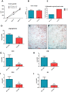
Figure 3. Inhibition of calpains in melanoma cells only: an in vivo approach.
C57BL/6 WT mice were injected with one million melanoma B16-F10 cells either transgenic for calpastatin (Calpast-Mel) or transfected with a control plasmid (Ctrl-Mel) and sacrificed at day 16 for tissues analysis. A. Tumor growth was measured from day 9 to day 16. Calpastatin overexpression in melanoma decreased significantly tumor growth. N = 10/group, * p< 0.05. B. Tumor weight at day 16. Calpastatin overexpression reduced significantly melanoma weight. N = 10/group, * p<0.05. C. Proportion of metastatic regional (axillary) lymph nodes at day 16. Mice injected with melanoma cells with reduced calpain activity (Calpast-Mel) had significantly more metastatic lymph nodes than controls (Ctrl-Mel) (10/10 vs 5/10 respectively, * p<0.05). D. Angiogenesis assessed by vessel count at 200×magnification after CD-31 (PECAM) staining. Neo-angiogenesis was similar in transgenic and control melanomas. N = 10/group, p = NS. E,F,G,H: CD3, CD4, CD8 and NK cell number/HPF (200×magnification). Immune cell infiltrate was similar in transgenic and control tumors. N = 10/group, p = NS.
Figure 4
Figure 4. Inhibition of calpains in melanoma cells only: an in vitro approach.
A. Cellular proliferation measured by BrdU incorporation (DNA synthesis). Calpastatin overexpression reduced significantly DNA synthesis under basal culture conditions. N = 4 independent experiments, * p<0.05. B. Quantification of apoptotic cells by Annexin V (+/- propidium iodide) staining by flow cytometry under basal conditions and after 24 hours exposure to mitomycin C. Melanoma cells overexpressing calpastatin were protected against mitomycin C-induced apoptosis. N = 5 independent experiments, # p<0.05 vs Ctrl-Mel+ Mitomycin C. C. Cytotoxic effect of previously immunized splenocytes (CTLs, previously immunized against control B16F10cells) from C57BL/6 mice (WT) or calpastatin transgenic mice (TG) against B16-F10 melanoma cells transfected with calpastatin (Calpast-Mel) or control plasmid (Ctrl-Mel). Cytotoxicity was measured by chromium release after incorporation in melanoma cells. TG CTLs and WT CTLs cells exerted a similar cytolytic response (p = NS) but Calpast-Mel cells were partly protected against immune effectors as compared to Ctrl-Mel cells. N = 4 independent experiments, * p<0.05. D, E. Melanoma cell migration measured by cell free gap surface recovery 10 hours after removing insert. Calpastatin overexpression increased significantly cellular migration properties in vitro. N = 6 independent experiments, * p<0.05.
Figure 5
Figure 5. Inhibition of calpains in host mice only.
C57BL/6 control (WT mice) or transgenic mice (CalpTG mice) were injected with one million melanoma B16F10 cells and sacrificed at day 16 for tissue analysis.A. Tumor growth was measured from day 11 to day 16. Calpastatin overexpression in host did not modify tumor growth. N = 10/group. B. Tumor weight at day 16. Calpastatin overexpression in hosts did not modify tumor weight. N = 10/group, p = NS. C. Proportion of metastatic regional lymph nodes at day 16. CalpTG mice had a trend to have more metastatic lymph nodes than WT mice (9/10 vs 4/10 respectively, N = 10/group, p = NS). D,E,F. Angiogenesis as assessed by vessel count at 200×magnification after CD-31 staining. Neo-angiogenesis was significantly decreased in CalpTG mice when compared to WT mice. N = 10/group, * p<0.05. G,H,I,J: CD3, CD4, CD8 and NK cell number/HPF (200×magnification). Immune cell infiltrate was significantly lower in CalpTG mice than in WT mice. N = 10/group, * p<0.05.
Figure 6
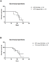
Figure 6. Survival studies.
A. Specific limitation of calpain activity in melanoma cells only by calpastatin overexpression in vivo. C57BL/6 WT mice were injected with one million melanoma B16-F10 cells either transgenic for calpastatin (Calpast-Mel) or transfected with a control plasmid (Ctrl-Mel), n = 10/group. Limitation of calpain activity in melanoma cells only did not modify survival at day 30. B. Specific limitation of calpain activity in mice transgenic for calpastatin. C57BL/6 control (WT mice) or transgenic mice (CalpTG mice) were injected with one million control melanoma B16F10 cells, n = 10/group. Limitation of calpain activity in host did not modify survival at day 30.

References

    1. Goll DE, Thompson VF, Li H, Wei W, Cong J (2003) The calpain system. Physiol Rev 83: 731–801. - PubMed
    1. Glading A, Uberall F, Keyse SM, Lauffenburger DA, Wells A (2001) Membrane proximal ERK signaling is required for M-calpain activation downstream of epidermal growth factor receptor signaling. J Biol Chem 276: 23341–8. - PubMed
    1. Su Y, Cui Z, Li Z, Block ER (2006) Calpain-2 regulation of VEGF-mediated angiogenesis. FASEB J 20: 1443–51. - PubMed
    1. Franco SJ, Huttenlocher A (2005) Regulating cell migration: calpains make the cut. J Cell Sci 118: 3829–38. - PubMed
    1. Dewitt S, Hallett M (2007) Leukocyte membrane "expansion": a central mechanism for leukocyte extravasation. J Leukoc Biol 81: 1160–4. - PubMed

Publication types