. 2013 Jan;2(1):1-15.
doi: 10.1002/wmts.72. Epub 2012 Dec 11.

Gap junctions and Bystander Effects: Good Samaritans and executioners

Affiliations

Gap junctions and Bystander Effects: Good Samaritans and executioners

David C Spray et al. Wiley Interdiscip Rev Membr Transp Signal. 2013 Jan.

Abstract

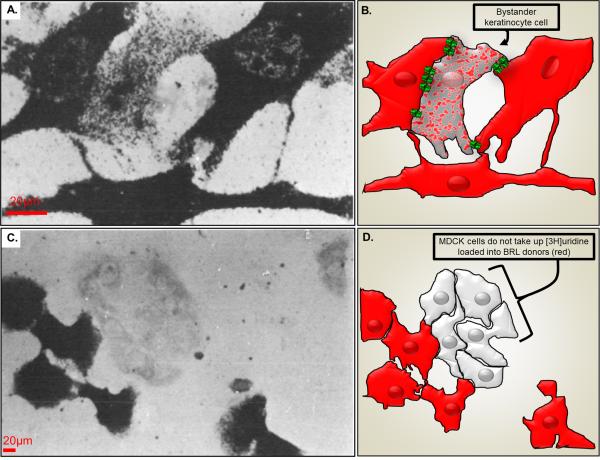
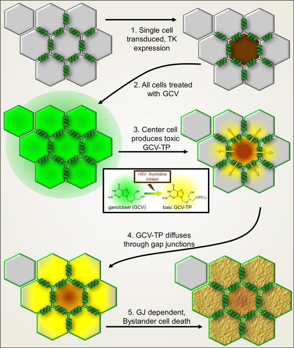
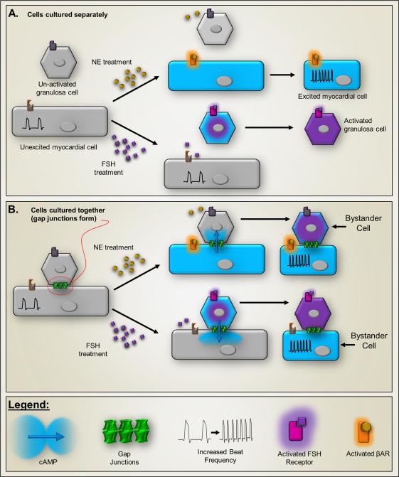
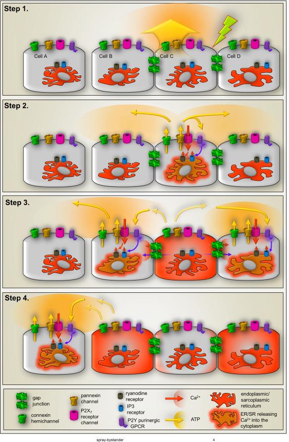
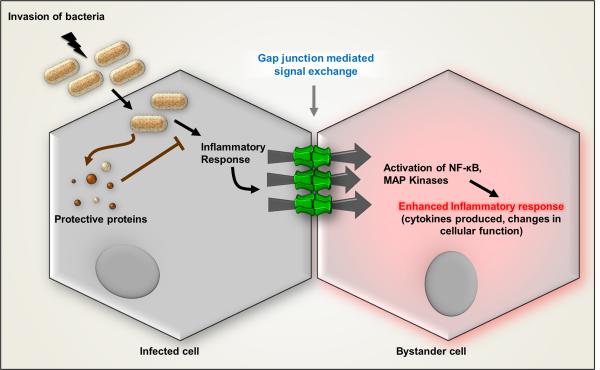
The "Bystander" and "Good Samaritan" effects involve the transfer of toxic or beneficial compounds from one cell to a generally adjacent other through gap junction channels and through extracellular routes. The variety of injuries in which bystander cell killing or protection occurs has greatly expanded in the last decade to include infectious agents and therapeutic compounds, radiation injury, chaperones in cell therapy and apoptosis in development. This has been accompanied by the appreciation that both gap junction mediated and paracrine routes are used for the signaling of the "kiss of life" and the "kiss of death" and that manipulations of these pathways and the molecules that use them may find therapeutic utility in treatment of a variety of pathological conditions.


Figures

Figure 1
Figure 1
Pioneering metabolic labeling experiments on coupling competent and coupling deficient cells. A.) Uridine nucleotide transfer between dermal fibroblast donors and epidermal keratinocyte recipients. Donor cell cultures were labeled with [3H]uridine and the autoradiograph was exposed for 3 weeks prior to developing. B.) Illustration showing keratinocyte (spotted cell) that has received [3H]uridine from the pre-loaded donor dermal fibroblast (red) likely through gap junctions (green). C.) Lack of uridine nucleotide transfer between BRL cells and MDCK cells. D.) Illustration of cell location where a lack of gap junction coupling leads to no [3H]uridine transfer from pre-loaded donor BRL cells (red) and MDCK cells (light gray). Modified from Hunter and Pitts.
Figure 2
Figure 2
Schematic diagram of Bystander cell killing as therapy. Cells interconnected by gap junction channels (one cell is not coupled by gap junctions). 1. Only one of eight cells is transduced by a virus with genetic code for the Herpes simplex virus thymidine kinase (HSV-tk), which induces expression in only that single cell (brown). 2. Treatment of the cell population with membrane permeant ganciclovir (GCV). 3. HSV-tk in the center cell converts GCV to the toxic phosphorylated and membrane impermeant GCV-triphosphate (GCV-TP, yellow color). 4. GCV-TP, with Mr 495 Da, is gap junction permeant and diffuses to neighboring cells. 5. Bystanders cells coupled to the center cell receive GCV-TP which intercalates into DNA, terminating synthesis and resulting in Bystander cell death.
Figure 3
Figure 3
Intercellular second messenger signaling evidenced in co-culture of mouse myocardial cells with rat ovarian granulosa cells. Myocardial cells respond to adrenergic agonists (βAR) with increased beat frequency (insets), whereas (B) granulosa cells produce plasminogen activator when stimulated by follicle stimulating hormone (FSH). In both cases, the second messenger is likely cAMP, which is elevated in the stimulated cell (illustrated by blue color). A.) Because the cell types express different receptors, when cultured alone neither responds to the other's agonist. B.) When cultured together myocardial and granulosa cells form gap junctions (green structures). Co-culture produces a Bystander effect in which addition of either norepinephrine or FSH to the co-cultures results in cross-stimulation, evoking changes in both cell types. This presumably results from cell-to-cell transfer of a second messenger, which is likely to be cyclic AMP (MW 329). Illustration represents experiments described in .
Fig. 4
Fig. 4
Components of intercellular calcium waves. Schematic representation of steps involved in transmission of intercellular Ca2+ waves through gap junctions and through ATP release. Step 1: Mechanical, electrical or pharmacological stimulation (green arrow) of one cell leads to Ca2+ entry through ion channels and ATP release (yellow arrow) that can occur through vesicular and non-vesicular mechanisms that involve transporters and channels such as pannexin1 channels and connexin hemichannels. Step 2: Autocrine activation of cell surface metabotropic P2Y (purple) and ionotropic P2X (magenta) receptors by released ATP induces increase in intracellular Ca2+ levels of the stimulated cell by Ca2+ influx (orange arrow) through P2XRs and Ca2+ mobilization from intracellular stores triggered by activation of IP3 receptors (blue cylinder) on endoplasmic/sarcoplasmic reticulum mediated by P2YR-generated IP3 (purple arrow) and Ca2+-induced Ca2+ release (small orange arrow) mediated by activation of ryanodine receptors (brown cylinder). Besides contributing to ATP-mediated Ca2+ influx, activation of the P2X7R subtype (magenta) also provide a mechanism for ATP-induced ATP release through its interaction and activation of pannexin1 channels (yellow. Opening of connexin hemichannels (green on cell surface) have also been proposed to contribute a pathway for ATP release from stimulated cells. Step 3: The Ca2+ signal generated in the stimulated cell spreads to adjacent cells when Ca2+ itself and the second messenger IP3 pass though gap junctions (green channels between cells), thereby inducing Ca2+ release from Ca2+ and IP3 sensitive intracellular stores (see Step 2), and when ATP activates P2 receptors in the adjacent cells (as in Step 2). Besides contributing to increase in intracellular Ca2+ levels of neighboring cells, paracrine activation of P2 receptors can induce further ATP release in the path of the Ca2+ wave spread through P2X7R-mediated activation of pannexin1 channels (as in Step 2). Step 4: Cells that are not coupled by gap junctions can be recruited into the Ca2+ wave spread and the Ca2+ signal can “jump” cell free spaces when released ATP diffuses and activates P2 receptors in these cells, triggering the events described in steps 2-3. Similar mechanisms contribute to long range signal spread.
Fig. 5
Fig. 5
Contagious Bystander. Bacterial infection generates proinflammatory signals in the infected cells that spread to the uninfected Bystander cells through gap junction channels to amplify the innate immune response. NF-kB and MAP kinases are activated in uninfected Bystanders to produce IL-8 following infection of adjacent cells. Modified from schematic diagram in Kasper et al..
Fig. 6
Fig. 6
Bystander cell death in the developing retina. a, b, Z-series projection (20 μm) of retinal sections from P5 mouse injected with vehicle and scrape-loaded with RD (rhodamine dextran) and NB (neurobiotin). a, RD labels cells in ganglion cell layer (GCL) and neuroblast layer (NBL, arrows). b, NB (reacted with Cy2-streptavidin) is localized in cells that were scrape-loaded with RD (arrows) and other cells not loaded with RD (arrowheads). Many processes in the inner plexiform layer (IPL) were labeled with NB (double arrowhead). Arrowheads in a indicate position of coupled cells in b, c, d, Carbenoxolone treatment decreases spread of NB. c, RD labeling (arrows) of scrape-loaded retina from animal treated with the gap junction blocker carbenoxolone (20 mg/kg, s.c.). d, NB labeling of same field shows only a few NB-labeled cells that do not colocalize with RD (arrowheads). Processes in IPL and pOPL are indicated by double arrowheads. Scale bars in a-d, 20 μm. e, Coupling Index for control (Con) and carbenoxolone (CBX) indicates a significant decrease in dye coupling by carbenoxolone treatment (asterisks indicate groups significantly different from controls; Student's t test, p < 0.01). f, carbenoxolone reduces the percentage of dying cells with a NN distance shorter than 15 μm in a manner that closely parallels the decrease in the Coupling Index. From Cusato et al,.
Fig. 7
Fig. 7
Cytochrome c (cyC) injection causes primary and Bystander cell death in Xeonpus oocytes. (ad) Time lapse photographs of electrically coupled oocytes following injection of cyC into the cell at the upper right. (a) At the time of injection, both cells have normal pigmentation and morphology. (b) At 30 min after introducing cyC, the injected cell shows a loss of pigmentation at the animal pole and has begun to swell. (c) At 60 min post-cyC, the injected cell continues to swell and becomes somewhat distorted. (d) At 120 min post-cyC, the injected cell has lost structural integrity and the uninjected ‘Bystander’cell has begun to lose pigmentation and swell. Gap junctional coupling measurements. (e) Protocol for dual cell two-electrode voltage clamp studies. Cells were held at -40 mV and stepped to -60 mV for 1 s. The downward deflection of the stepped cell is the total current (Gtotal), which is the sum of the junctional current (Gj) and nonjunctional current (Gnj). The upward deflection in the follower cell is the current that has passed from one cell to the other via gap junctions (Gj). (f) Current recordings from a pair of cells in which one cell was injected with cyC at t=0 min. The Gj and Gnj in the cyC-injected cell (I cyC) increase as the cell dies. The Bystander cell also undergoes an increase in Gj equal to that of the cyC-injected cell (I B), but the increase in Gnj is less pronounced. Bottom two traces show the timing of the voltage pulses to each cell, which were 1 s in duration and 10 s apart. From Cusato et al..

References

    1. Pitts JD. The discovery of metabolic co-operation. Bioessays. 1998;20:1047–51. - PubMed
    1. Fujimoto WY, Subak-Sharpe JH, Seegmiller JE. Hypoxanthine-guanine phosphoribosyltransferase deficiency: chemical agents selective for mutant or normal cultured fibroblasts in mixed and heterozygote cultures. Proc Natl Acad Sci U S A. 1971;68:1516–9. - PMC - PubMed
    1. Kimura S, Hase K, Ohno H. Tunneling nanotubes: emerging view of their molecular components and formation mechanisms. Experimental cell research. 2012;318:1699–706. - PubMed
    1. Hamada N, Maeda M, Otsuka K, Tomita M. Signaling pathways underpinning the manifestations of ionizing radiation-induced bystander effects. Curr Mol Pharmacol. 2011;4:79–95. - PubMed
    1. Yang L, Cranson D, Trinkaus-Randall V. Cellular injury induces activation of MAPK via P2Y receptors. J Cell Biochem. 2004;91:938–50. - PubMed

LinkOut - more resources