. 2013 Apr;5(2):131-46.
doi: 10.2217/epi.13.1.

Epigenetic regulation of Sox4 during palate development

Affiliations

Epigenetic regulation of Sox4 during palate development

Ratnam S Seelan et al. Epigenomics. 2013 Apr.

Abstract

Aim: Identification of genes that contribute to secondary palate development provide a better understanding of the etiology of palatal clefts. Gene-expression profiling of the murine palate from gestational days 12-14 (GD12-14), a critical period in palate development, identified Sox4 as a differentially expressed gene. In this study, we have examined if the differential expression of Sox4 in the palate is due to changes in DNA methylation.

Materials & methods: In situ hybridization analysis was used to localize the expression of Sox4 in the developing murine secondary palate. CpG methylation profiling of a 1.8-kb upstream region of Sox4 in the secondary palate from GD12-14 and transfection analysis in murine embryonic maxillary mesenchymal cells using Sox4 deletion, mutant and in vitro methylated plasmid constructs were used to identify critical CpG residues regulating Sox4 expression in the palate.

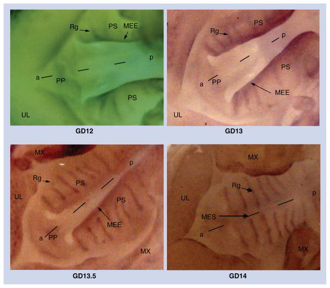
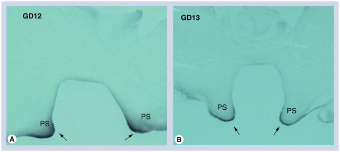
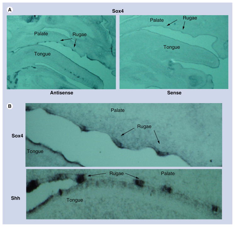
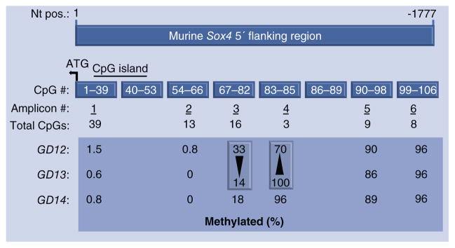
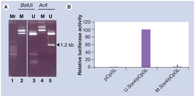
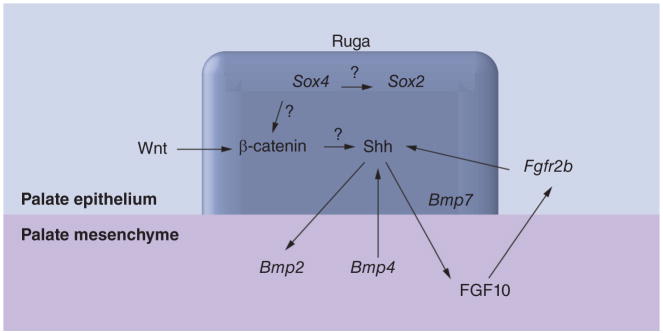
Results: Spatiotemporal analysis revealed that Sox4 is expressed in the medial edge epithelium and presumptive rugae-forming regions of the palate from GD12 to GD13. Following palatal shelf fusion on GD14, Sox4 was expressed exclusively in the epithelia of the palatal rugae, structures that serve as signaling centers for the anteroposterior extension of the palate, and that are thought to serve as neural stem cell niches. Methylation of a 1.8-kb region upstream of Sox4, containing the putative promoter, completely eliminated promoter activity. CpG methylation profiling of the 1.8-kb region identified a CpG-poor region (DMR4) that exhibited significant differential methylation during palate development, consistent with changes in Sox4 mRNA expression. Changes in the methylation of DMR4 were attributed primarily to CpGs 83 and 85.

Conclusion: Our studies indicate that Sox4 is an epigenetically regulated gene that likely integrates multiple signaling systems for mediating palatal fusion, palatal extension and/or the maintenance of the neural stem cell niche in the rugae.


Figures

Figure 1
Figure 1. Whole-mount in situ hybridization with Sox4 riboprobes on GD12–GD14 palatal processes
Sox4 expression in the developing murine secondary palate was determined from GD12 to GD14 using antisense riboprobes. On GD12 and GD13, Sox4 expression is visible in the MEE and the developing rugae. At GD13.5, Sox4 expression is more distinct in the rugae and expression persists at the medial edge epithelium. After palatal fusion on GD14, expression is confined only to the rugae with no evidence of expression at the MES where the palatal shelves have fused. Representative photomicrographs depict ventral views of the oral region of GD12–GD14 mouse embryos subsequent to removal of the mandible and tongue. a–p: Anteroposterior axis; MEE: Medial edge epithelium; MES: Medial edge seam; MX: Maxilla; PP: Primary palate; PS: Palatal shelf; Rg: Ruga; UL: Upper lip.
Figure 2
Figure 2. Sectional in situ hybridization with Sox4 riboprobes on coronal sections of GD12 and GD13 palatal processes
Coronal sections of secondary palates from (A) GD12 and (B) GD13 mice were hybridized with Sox4 antisense riboprobes. Representative photomicrographs indicate intense Sox4 expression in the inferior margins of vertically directed palatal processes (presumptive medial edge epithelium) on both GD12 and GD13 PS (arrows). PS: Palatal shelf.
Figure 3
Figure 3. Sectional in situ hybridization with Shh and Sox4 riboprobes on sagittal sections of GD14 oral/facial region and palatal processes
(A) Sagittal sections of oral/facial region from GD14 mice probed with Sox4 sense and antisense riboprobes. Representative photomicrographs indicate that Sox4 expression, in the secondary palate, is specifically localized to the rugae of the palate on GD14. (B) Sagittal sections of secondary palates from GD14 mice probed with Sox4 and Shh antisense riboprobes. Similar to Shh (which is expressed in the rugae epithelium to direct anteroposterior extension of the palate), Sox4 is also found to be expressed in the epithelial thickenings of the palatal rugae.
Figure 4
Figure 4. CpG methylation profiling of the 5′ upstream region of Sox4 in the developing murine secondary palate
A 1777 nt region upstream of the translational start site (+1) of murine Sox4 (accession number: NC_000079.6, NCBI) was used for CpG methylation profiling. After ‘bisulfite’ treatment of genomic DNA, six PCR amplicons were generated, spanning the 5′ upstream region. The 1777 nt region contained a total of 106 CpG residues, numbered sequentially from nt +1. Amplicon 1 represents the most proximal region and includes CpGs 1–39. A CpG island (horizontal bar) spans CpG residues 13–51. Regions containing CpGs 40–53 and 86–89 were not amplified (see text). The large box compares CpG methylation levels (%) in genomic DNA derived from secondary palates on GD12, GD13 and GD14. The two vertical boxes indicate the two amplicons exhibiting the most pronounced changes in methylation levels between GD12 and GD13 (note that methylation levels between GD13 and GD14 for these two amplicons are almost identical). Notably, amplicon 3 exhibited a decrease in methylation (33–14%) and amplicon 4 exhibited an increase (70–100%). The increase in methylation in amplicon 4 was consistent with decreasing mRNA expression observed between GD12 and GD13.
Figure 5
Figure 5. Functional analysis of CpG83 and CpG85 on Sox4 promoter activity
Various Sox4 constructs in luciferase reporter vectors were transfected into murine embryonic maxillary mesenchymal cells. (A) Values in the y-axis represent normalized luciferase activities. The Sox4 reporter activities of all constructs were compared with that of the SV40 promoter (column #2; taken as 100%). Plasmids used for transfection included: (#1) pGL3 vector; (#2) SV40/pGL3; (#3) Sox4/pGL3; (#4) Sox4Δ/pGL3; (#5) Sox4Mut83/pGL3; and 6) Sox4Mut85/pGL3. Note the significant decrease in promoter activities of the last three constructs (#4–#6) when compared with that of Sox4/pGL3 (#3). (B) Indicates the salient features of the Sox4 plasmid constructs utilized. The location of the two differentially methylated regions and the CpG island is indicated in Sox4/pGL3, which harbors the 1.8-kb 5′ upstream region of Sox4. X denotes mutated CpG sites. Sox4/pCpGL was used for methylation analysis and harbors the 1.8-kb 5′ upstream region of Sox4 (only CpGs 83 and 85 are indicated in the figure). Transfections were repeated four times using duplicates and values are presented as mean ± standard deviation. *p< 0.0001; **p < 0.0002. DMR: Differentially methylated region; Mut: Mutation; SV: Simian virus 40.
Figure 6
Figure 6. Effect of methylation of the 5′ upstream region on Sox4 promoter activity
(A) Sox4/pCpGL harboring the 1.8-kb 5′ upstream region of Sox4 (see Figure 5B) was methylated in vitro using SssI methylase. Methylation of the plasmid was assessed by digesting the unmethylated and methylated plasmids with BstUI and AclI. The methylated plasmids were resistant to digestion (lanes 2 and 4), whereas the unmethylated plasmids (lanes 3 and 5) released expected digestion products. CpG85 is one of only two sites in Sox4/pCpGL that can be digested by AclI. The figure clearly indicates that CpG85 is fully methylated, as evidenced by the absence of the 1.2-kb digestion product from the methylated plasmid (lane 4) when compared with that derived from digestion of the unmethylated plasmid (lane 5). (B) Normalized promoter activities of the methylated and unmethylated plasmids after transfection into murine embryonic maxillary mesenchymal cells are presented. The relative promoter activity of the unmethylated plasmid was set at 100%. The experiment was repeated four times using duplicates and values are presented as mean ± standard deviation (*p < 0.0001). M: Methylated; Mr: Marker; U: Unmethylated.
Figure 7
Figure 7. A model for SOX4 regulation in the rugae epithelium during secondary palate development
A model illustrating how SOX4 potentially integrates diverse developmental signals in the rugae during secondary palate development is presented. The ruga (boxed) is the Shh signaling center. Shh signaling facilitates palate development through epithelial–mesenchymal cross-talk involving Wnt, FGF and BMP signaling pathways. SOX4 acts to increase the stability of β-catenin, thereby acting as a Wnt agonist, and/or activates SOX2, a pluripotent factor, via binding to the Sox2 enhancer. The latter is consistent with the rugae being a niche for neural stem cells. ‘?’ denotes interactions whose underlying mechanisms are unknown. All other interactions are from published literature [,,–61].

References

    1. Yazdy MM, Honein MA, Rasmussen SA, Frias J. Priorities for future public health research in orofacial clefts. Cleft Palate Craniofac J. 2007;44(4):351–357. - PubMed
    1. Gritli-Linde A. Molecular control of secondary palate development. Dev Biol. 2007;301(2):309–326. - PubMed
    1. Juriloff DM, Harris MJ. Mouse genetic models of cleft lip with or without cleft palate. Birth Defects Res A Clin Mol Teratol. 2008;82(2):63–77. Comprehensive review that identifies Sox4 as a candidate gene for human nonsyndromic cleft lip with cleft palate. - PubMed
    1. Lidral AC, Moreno LM. Progress toward discerning the genetics of cleft lip. Curr Opin Pediatr. 2005;17(6):731–739. - PMC - PubMed
    1. Carinci F, Scapoli L, Palmieri A, Zollino I, Pezzetti F. Human genetic factors in nonsyndromic cleft lip and palate: an update. Int J Pediatr Otorhinolaryngol. 2007;71(10):1509–1519. - PubMed

Websites

    1. EMBOSS explorer cpgplot. www.bioinformatics.nl/cgi-bin/emboss/cpgplot

    1. gene-regulation.com. www.gene-regulation.com/pub/programs.html

Publication types

Substances

LinkOut - more resources