. 2013 Jul;34(21):4971-81.
doi: 10.1016/j.biomaterials.2013.03.015. Epub 2013 Apr 6.

Modeling vascularized bone regeneration within a porous biodegradable CaP scaffold loaded with growth factors

Affiliations

Modeling vascularized bone regeneration within a porous biodegradable CaP scaffold loaded with growth factors

Xiaoqiang Sun et al. Biomaterials. 2013 Jul.

Abstract

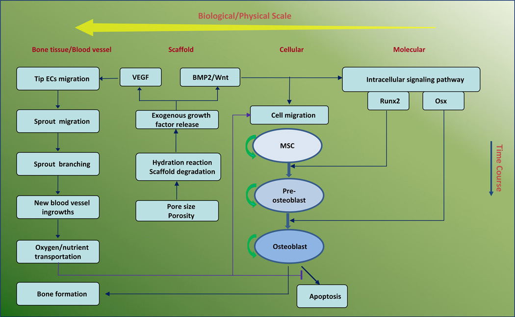
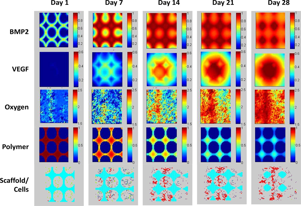
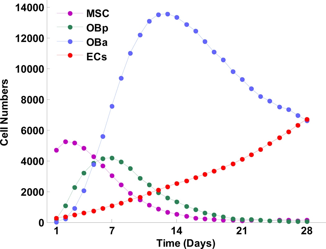
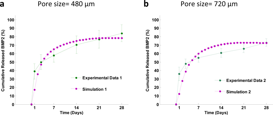
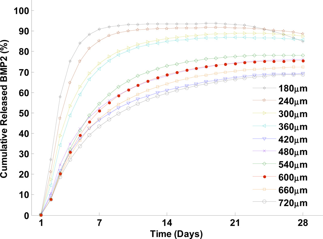
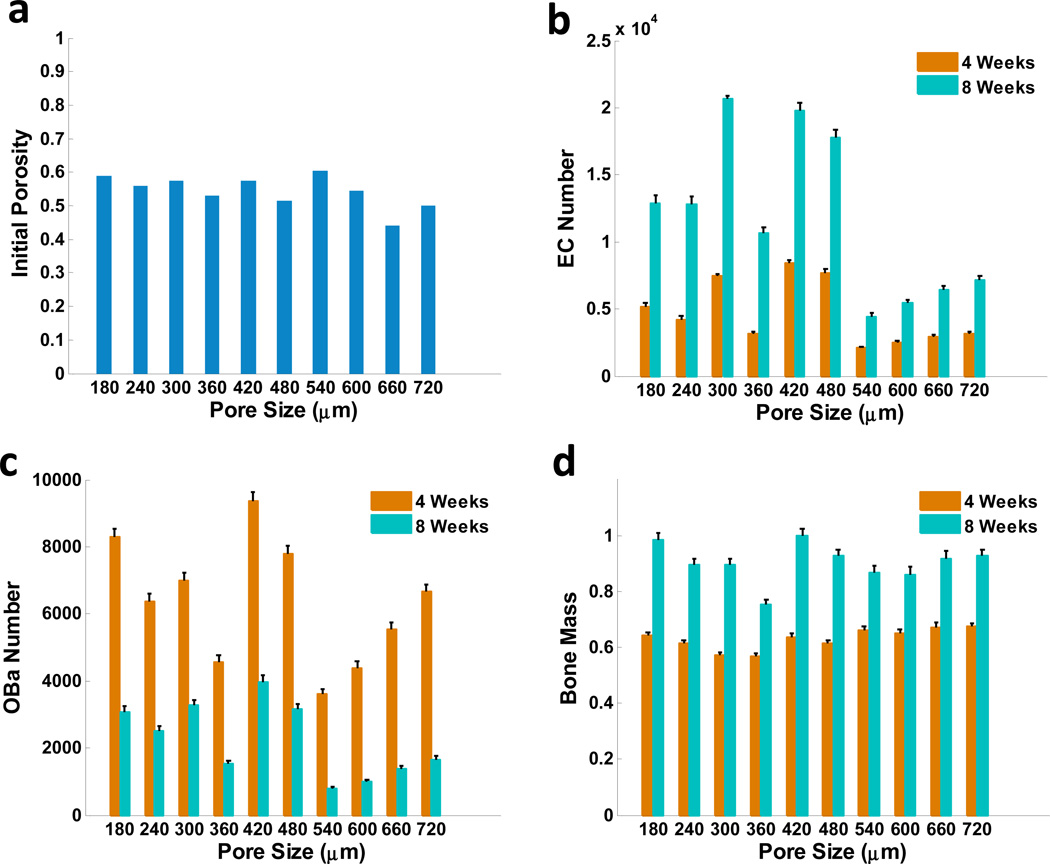
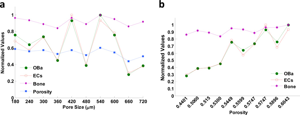
Osteogenetic microenvironment is a complex constitution in which extracellular matrix (ECM) molecules, stem cells and growth factors each interact to direct the coordinate regulation of bone tissue development. Importantly, angiogenesis improvement and revascularization are critical for osteogenesis during bone tissue regeneration processes. In this study, we developed a three-dimensional (3D) multi-scale system model to study cell response to growth factors released from a 3D biodegradable porous calcium phosphate (CaP) scaffold. Our model reconstructed the 3D bone regeneration system and examined the effects of pore size and porosity on bone formation and angiogenesis. The results suggested that scaffold porosity played a more dominant role in affecting bone formation and angiogenesis compared with pore size, while the pore size could be controlled to tailor the growth factor release rate and release fraction. Furthermore, a combination of gradient VEGF with BMP2 and Wnt released from the multi-layer scaffold promoted angiogenesis and bone formation more readily than single growth factors. These results demonstrated that the developed model can be potentially applied to predict vascularized bone regeneration with specific scaffold and growth factors.


Figures

Fig. 1
Fig. 1
Schematic representation of computational framework of 3D vascularized bone regeneration within a porous biodegradable CaP scaffold. The model encapsulates four biological/physical scales from micro level to macro level: molecular, cellular, scaffold, and bone tissue scales. At the scaffold scale, growth factors (BMP2, Wnt and VEGF) were released from the CaP scaffold via calcium phosphate degradation due to hydration reaction. At molecular scale, the released BMP2 and Wnt stimulated the intracellular signaling pathway of MSC and pre-osteoblasts to activate the transcription factors Runx2 and Osx. At the cellular scale, each osteoblastic cell underwent migration, proliferation, differentiation, and apoptosis. MSC and pre-osteoblasts migrated along the gradient of the concentration of growth factors and oxygen, and their differentiation was regulated by Runx2 and Osx. At the bone tissue scale, new capillary sprouts, migrating from host tissue, were induced and sustained by released VEGF to grow into the pores of the scaffold. The remodelled vasculature could transport oxygen to maintain osteoblast metabolism and survival within scaffold pores.
Fig. 2
Fig. 2
A typical simulation from a 3D model showing the evolution of scaffold resorption, angiogenesis, MSC differentiation, and cell growth within scaffold pores over time. Different colors denote various cell types: yellow (MSC), green (pre-osteoblasts), blue (active osteoblasts), and red (blood vessels). The uniformly distributed pores on the scaffolds gradually degraded due to resorption of the scaffold. Newly formed blood vessels grew into the pores at the periphery of the scaffold.
Fig. 3
Fig. 3
2D cross-sectional view of the spatio-temporal evolution of BMP2 concentration, VEGF concentration, oxygen concentration, calcium phosphate molecular weight, and scaffold/cell profiles over time. BMP2 and VEGF were released from the calcium phosphate matrix, and continuously diffused into the scaffold pores. Oxygen concentrations changed because of uptake by osteoblasts and transportation by the neovasculature. Calcium phosphate molecular weight decreased because of hydrolysis. Bottom row: degradation of porous CaP scaffold (cyan) and the differentiation of MSC (yellow) into active osteoblasts (blue), as well as angiogenesis (red).
Fig. 4
Fig. 4
Predicted changes in numbers of cell types over time. Peaks of different cell types occurred at different times, reflecting osteoblast phenotype development through the osteoblast lineage. ECs, endothelial cells; MSC, mesenchymal stem cells; OBa, active osteoblasts; and OBp, pre-osteoblasts.
Fig. 5
Fig. 5
Predictions of cumulative released BMP2 from scaffolds with (a) small pore size (480 µm) and (b) big pore size (720 µm), compared with our experimental data.
Fig. 6
Fig. 6
Predictions of cumulative released BMP2 from scaffolds (measured by the concentration of released BMP2 in the pore space) as a function of different pore sizes.
Fig. 7
Fig. 7
Predicted effects of scaffold porosity and pore size on angiogenesis and bone formation. (a) Initial porosities of scaffolds with different pore sizes. (b) Normalized numbers of endothelial cells (c) active osteoblasts; and (d) bone mass as a function of different pore sizes after 4 and 8 weeks, respectively.
Fig. 8
Fig. 8
The correlations between the scaffold porosity or pore size and the number of active osteoblasts, endothelial cells, and bone mass. (a) After 8 weeks, predicted numbers of active osteoblasts and endothelial cells, and bone mass formation showed a greater correlation to scaffold porosity than to scaffold pore sizes. (b) Numbers of active osteoblasts (OBa) and endothelial cells (ECs), as well as bone mass formation, were strongly related to porosity. "Normalized values" indicates that these values were normalized to their original maximum values.
Fig. 9
Fig. 9
Growth factor combination prediction. In therapy case a, 10 dose of Wnt was used; in therapy case b, 10 dose of BMP2 was used; in therapy case c, a combination of 4 dose of Wnt, 2 dose of BMP2 and 4 dose of VEGF were used; in therapy case d, a combination of 2 dose of Wnt, 4 dose of BMP2 and 4 dose of VEGF were used. (a) The evolution of the numbers of active osteoblasts from day 1 to day 56 under different therapies. (b) The normalized number of endothelial cells under different therapies evaluated in 4th and 8th week. (c) The normalized bone mass under different therapies evaluated in 4th and 8th week. The most efficient growth of active osteoblasts was promoted by the combination of Wnt, BMP2, and VEGF (case c and d). The number of endothelial cells increased dramatically in therapies with VEGF (case c and d) compared with therapies without VEGF (case a and b). More bone formation was observed after 8 weeks when different growth factors were combined together (case c and d) than the therapies of single growth factor (case a and b).

References

    1. Petite H, Viateau V, Bensaid W, Meunier A, De Pollak C, Bourguignon M, et al. Tissue-engineered bone regeneration. Nature Biotechnol. 2000;18:959–963. - PubMed
    1. Dragoo JL, Lieberman JR, Lee RS, Deugarte DA, Lee Y, Zuk PA, et al. Tissue-engineered bone from BMP-2--transduced stem cells derived from human fat. Plast Reconstr Surg. 2005;115:1665–1673. - PubMed
    1. Trautvetter W, Kaps C, Schmelzeisen R, Sauerbier S, Sittinger M. Tissue-engineered polymer-based periosteal bone grafts for maxillary sinus augmentation: five-year clinical results. J Oral Maxillofac Surg. 2011;69:2753–2762. - PubMed
    1. Nguyen LH, Annabi N, Nikkhah M, Bae H, Binan L, Park S, et al. Vascularized bone tissue engineering: approaches for potential improvement. Tissue Eng Part B Rev. 2009;15:363–382. - PMC - PubMed
    1. Lovett M, Lee K, Edwards A, Kaplan DL. Vascularization strategies for tissue engineering. Tissue Eng Part B Rev. 2009;15:353–370. - PMC - PubMed

Publication types

MeSH terms

Substances