

Patterns of reduced cortical thickness in late-life depression and relationship to psychotherapeutic response
- PMID: 23567394
- PMCID: PMC3732520
- DOI: 10.1016/j.jagp.2013.01.013
Patterns of reduced cortical thickness in late-life depression and relationship to psychotherapeutic response
Abstract
Objective: Cortical atrophy has been associated with late-life depression (LLD) and recent findings suggest that reduced right hemisphere cortical thickness is associated with familial risk for major depressive disorder, but cortical thickness abnormalities in LLD have not been explored. Furthermore, cortical atrophy has been posited as a contributor to poor antidepressant treatment response in LLD, but the impact of cortical thickness on psychotherapy response is unknown. This study was conducted to evaluate patterns of cortical thickness in LLD and in relation to psychotherapy treatment outcomes.
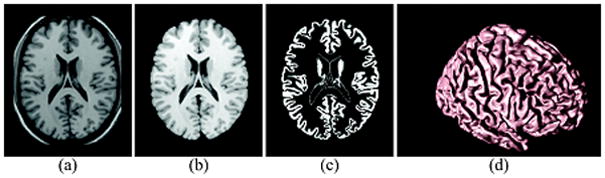
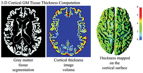
Methods: Participants included 22 individuals with LLD and 12 age-matched comparison subjects. LLD participants completed 12 weeks of psychotherapy and treatment response was defined as a 50% reduction in depressive symptoms. All participants underwent magnetic resonance imaging of the brain, and cortical mapping of gray matter tissue thickness was calculated.
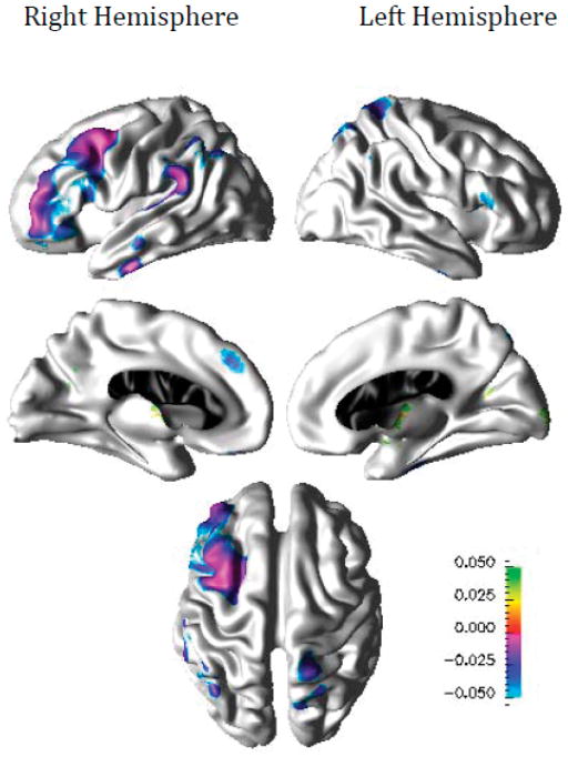
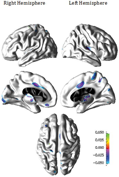
Results: LLD individuals demonstrated thinner cortex than controls prominently in the right frontal, parietal, and temporal brain regions. Eleven participants (50%) exhibited positive psychotherapy response after 12 weeks of treatment. Psychotherapy nonresponders demonstrated thinner cortex in bilateral posterior cingulate and parahippocampal cortices, left paracentral, precuneus, cuneus, and insular cortices, and the right medial orbitofrontal and lateral occipital cortices relative to treatment responders.
Conclusions: Our findings suggest more distributed right hemisphere cortical abnormalities in LLD than have been previously reported. In addition, our findings suggest that reduced bilateral cortical thickness may be an important phenotypic marker of individuals at higher risk for poor response to psychotherapy.
Keywords: Atrophy; MRI; cortical thickness; geriatric; late-life depression; psychotherapy.
Copyright © 2013 American Association for Geriatric Psychiatry. Published by Elsevier Inc. All rights reserved.
Conflict of interest statement
There are no financial or other relationships that could be interpreted as a conflict of interest affecting this manuscript.
Figures
References
-
- Hasin DS, Goodwin RD, Stinson FS, et al. Epidemiology of major depressive disorder: results from the National Epidemiologic Survey on Alcoholism and Related Conditions. Arch Gen Psychiatry. 2005;62:1097–1106. - PubMed
-
- Anstey KJ, von Sanden C, Sargent-Cox K, et al. Prevalence and risk factors for depression in a longitudinal, population-based study including individuals in the community and residential care. Am J Geriatr Psychiatry. 2007;15:497–505. - PubMed
-
- Sneed JR, Kasen S, Cohen P. Early-life risk factors for late-onset depression. Int J Geriatr Psychiatry. 2007;22:663–667. - PubMed
-
- Holley C, Murrell SA, Mast BT. Psychosocial and vascular risk factors for depression in the elderly. Am J Geriatr Psychiatry. 2006;14:84–90. - PubMed
-
- Egger K, Schocke M, Weiss E, et al. Pattern of brain atrophy in elderly patients with depression revealed by voxel-based morphometry. Psychiatry Res. 2008;164:237–244. - PubMed
